Review
. 2022 Feb 6;11(3):568.
doi: 10.3390/cells11030568.

The Pivotal Role of the Placenta in Normal and Pathological Pregnancies: A Focus on Preeclampsia, Fetal Growth Restriction, and Maternal Chronic Venous Disease

Affiliations
Review

The Pivotal Role of the Placenta in Normal and Pathological Pregnancies: A Focus on Preeclampsia, Fetal Growth Restriction, and Maternal Chronic Venous Disease

Miguel A Ortega et al. Cells. .

Abstract

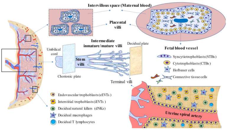
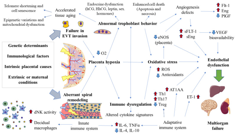
The placenta is a central structure in pregnancy and has pleiotropic functions. This organ grows incredibly rapidly during this period, acting as a mastermind behind different fetal and maternal processes. The relevance of the placenta extends far beyond the pregnancy, being crucial for fetal programming before birth. Having integrative knowledge of this maternofetal structure helps significantly in understanding the development of pregnancy either in a proper or pathophysiological context. Thus, the aim of this review is to summarize the main features of the placenta, with a special focus on its early development, cytoarchitecture, immunology, and functions in non-pathological conditions. In contraposition, the role of the placenta is examined in preeclampsia, a worrisome hypertensive disorder of pregnancy, in order to describe the pathophysiological implications of the placenta in this disease. Likewise, dysfunction of the placenta in fetal growth restriction, a major consequence of preeclampsia, is also discussed, emphasizing the potential clinical strategies derived. Finally, the emerging role of the placenta in maternal chronic venous disease either as a causative agent or as a consequence of the disease is equally treated.

Keywords: fetal growth restriction; maternal chronic venous disease (CVeD); placenta; preeclampsia.


Conflict of interest statement

The authors declare no conflict of interest.

Figures

Figure 1
Figure 1
An integrative picture of the components of the placenta. Herein, the main cell types and structures formed are summarized. At the top, placental villi and their cells, as well as the location of the maternal and fetal blood, in the intervillous space and inside the villi, respectively, are represented. In the center of the image, the villous tree, as well as the different types of villi, are represented. The chorionic plate, or fetal surface, is covered by the amnion, where the umbilical cord is attached. The decidual basal plate or maternal surface is in contact with the endometrium. As represented at the bottom, profound remodeling of the uterine spiral arteries is mainly due to the coordinated efforts of a set of cells, mainly extravillous trophoblasts (EVTs) and immune cells, prominently represented by decidual natural killers (dNKs). Having complete knowledge of the placenta in non-pathologic pregnancies is crucial for the study of different pregnancy complications, as the placenta is responsible for a wide variety of functions, as is subsequently discussed.
Figure 2
Figure 2
An overview of the main mechanisms involved in the pathogenesis of early-onset preeclampsia. As described before, aberrant spiral artery remodeling and failures in trophoblast invasion trigger placental hypoxia, leading to an exacerbated immune response affecting the innate and adaptative immune systems and cytokine production. In turn, an abnormal trophoblast behavior is also observed in the placenta, including accelerated aging, endocrine dysfunction, enhanced cell death, and angiogenesis defects, emphasizing the relevance of an imbalance between anti-angiogenic (sflt-1 and sEng) and pro-angiogenic factors (VEGF and PlGF). Almost every cell type in the placenta increases ROS production and decreases antioxidants, which are associated with oxidative stress, affecting some crucial components in the placenta, such as endothelial nitric oxide synthase, with detrimental effects on angiogenesis. Overall, oxidative stress, trophoblast alterations, and the inflammatory environment lead to systemic and profound endothelial dysfunction, eventually affecting different organs in the body and leading to the onset of clinical manifestations.
Figure 3
Figure 3
The vital role of the placenta in fetal growth restriction, risk factors, and consequences of the impaired maternofetal circulation. Main diagnosis, screening, and monitoring methods, as well as some preventive measures and recommendations, are also described.

Similar articles

Cited by

References

    1. Gude N.M., Roberts C.T., Kalionis B., King R.G. Growth and function of the normal human placenta. Thromb. Res. 2004;114:397–407. doi: 10.1016/j.thromres.2004.06.038. - DOI - PubMed
    1. Burton G.J., Jauniaux E. What is the placenta? Am. J. Obstet. Gynecol. 2015;213:S6.e1–S6.e4. doi: 10.1016/j.ajog.2015.07.050. - DOI - PubMed
    1. Huppertz B. Placental pathology in pregnancy complications. Thromb. Res. 2011;127:S96–S99. doi: 10.1016/S0049-3848(11)70026-3. - DOI - PubMed
    1. Konkel L. Lasting Impact of an Ephemeral Organ: The Role of the Placenta in Fetal Programming. Environ. Health Perspect. 2016;124:A124–A129. doi: 10.1289/ehp.124-A124. - DOI - PMC - PubMed
    1. Thornburg K.L., Marshall N. The placenta is the center of the chronic disease universe. Am. J. Obstet. Gynecol. 2015;213:S14–S20. doi: 10.1016/j.ajog.2015.08.030. - DOI - PMC - PubMed

Publication types