Review
. 2022 Feb 6;11(3):570.
doi: 10.3390/cells11030570.

Carboxypeptidase A3-A Key Component of the Protease Phenotype of Mast Cells

Affiliations
Review

Carboxypeptidase A3-A Key Component of the Protease Phenotype of Mast Cells

Dmitri Atiakshin et al. Cells. .

Abstract

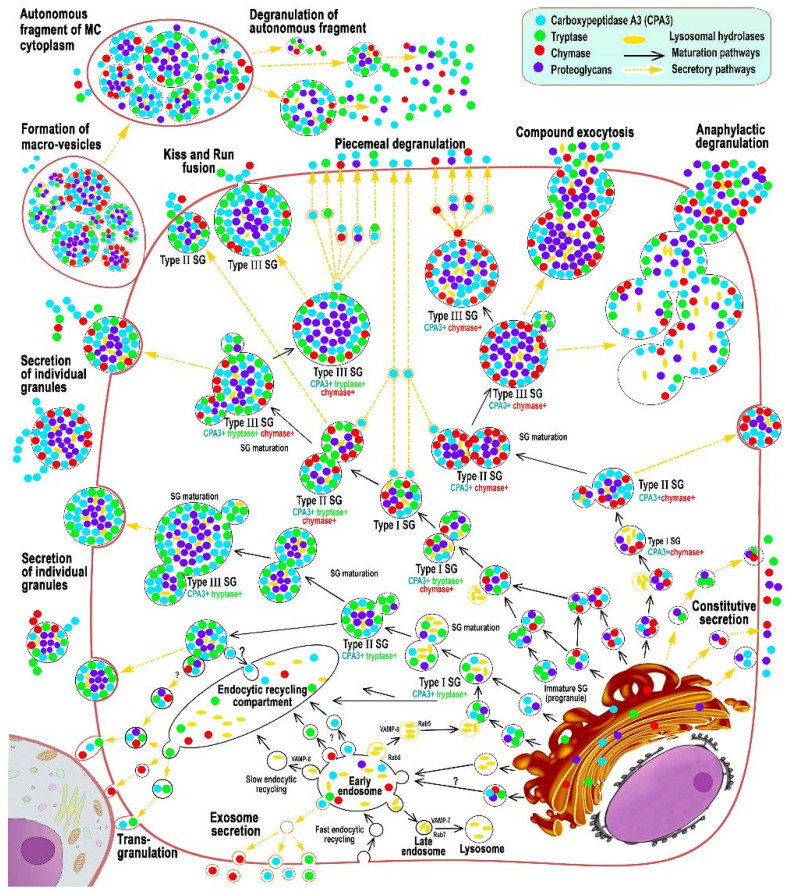
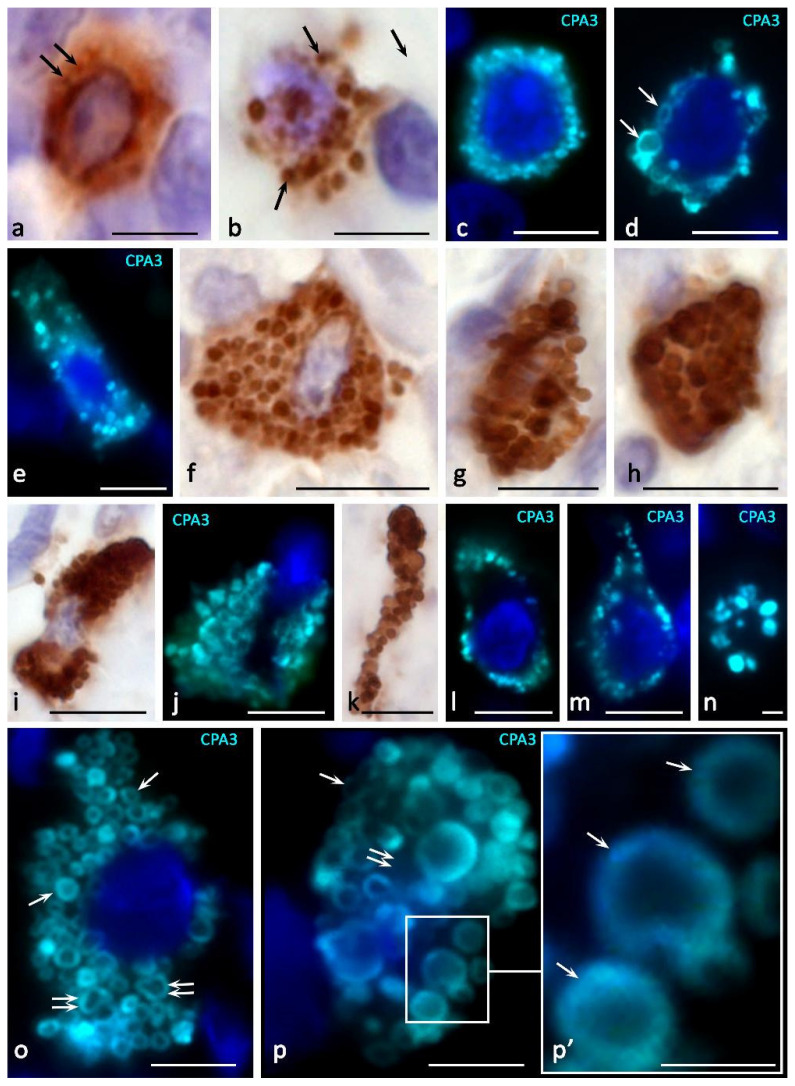
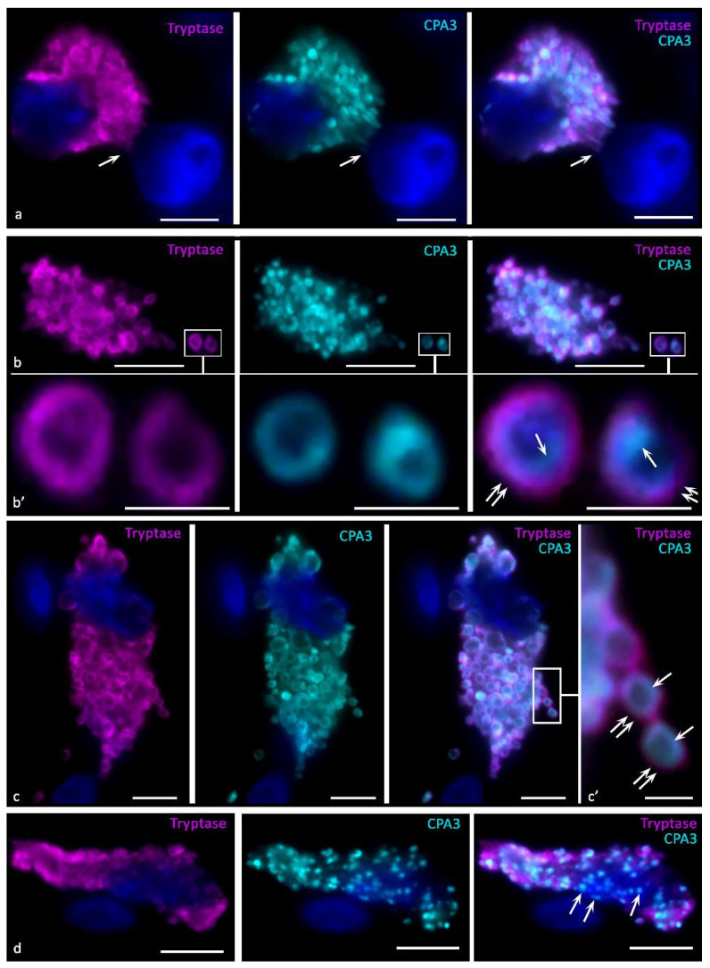
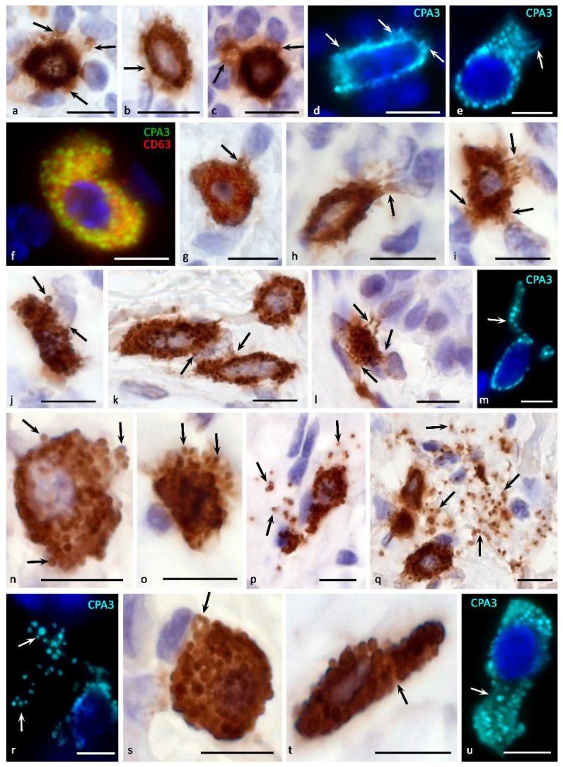
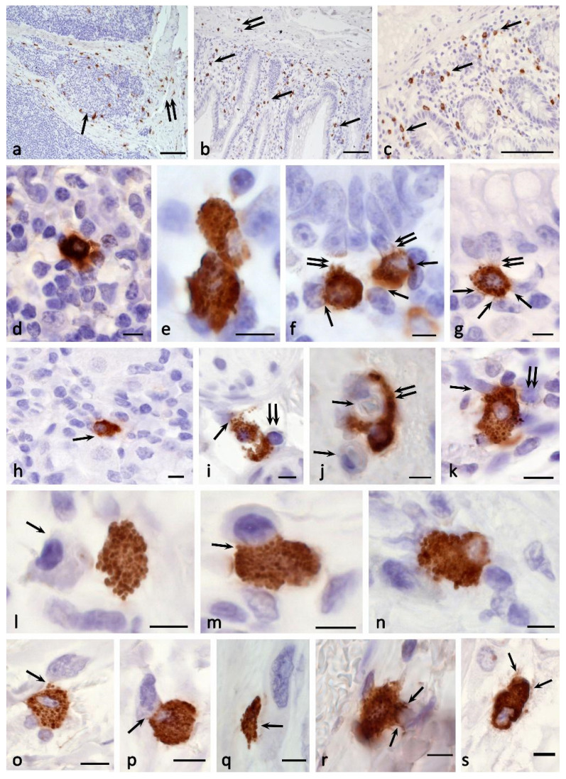
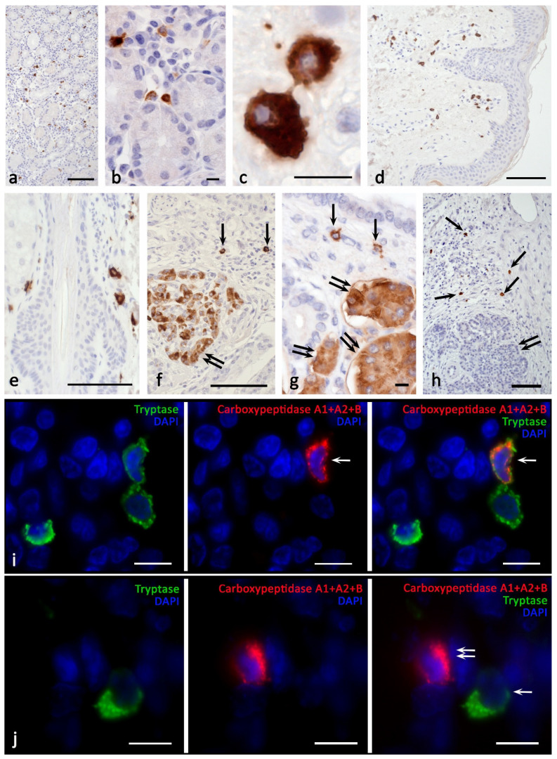
Carboxypeptidase A3 (CPA3) is a specific mast cell (MC) protease with variable expression. This protease is one of the preformed components of the secretome. During maturation of granules, CPA3 becomes an active enzyme with a characteristic localization determining the features of the cytological and ultrastructural phenotype of MC. CPA3 takes part in the regulation of a specific tissue microenvironment, affecting the implementation of innate immunity, the mechanisms of angiogenesis, the processes of remodeling of the extracellular matrix, etc. Characterization of CPA3 expression in MC can be used to refine the MC classification, help in a prognosis, and increase the effectiveness of targeted therapy.

Keywords: carboxypeptidase A3; granules; mast cells; secretome; secretory pathways; specific tissue microenvironment.


Conflict of interest statement

The authors declare no competing interests.

Figures

Figure 1
Figure 1
Main stages of biogenesis and secretory pathways of mast cell carboxypeptidase A3 (adapted from Atiakshin [84]). The diagram shows the main stages of post-translational modification of CPA3 with an emphasis on cytotopography, intragranular localization, and secretory mechanisms. CPA3 biosynthesis begins in the granular endoplasmic reticulum mast cells (MC) and continues in the Golgi apparatus (GA), where the progranule is formed. Secretory granules (SG) can be classified into 3 types. Type I SGs are formed after fusion of lysosomes with progranules, budding from GA, and have the smallest size and initial CPA3 content. The size of type I granules can increase due to homotypic fusion with the formation of type II granules with a size of 0.2–0.4 μm. Type II secretory granules already possess a certain phenotype of specific proteases. As a result of the completion of maturation, type III SGs are formed with a size of 0.5 μm and more, which are characterized by the largest volume of secretome and peripheral localization of CPA3 in the form of a ring. The main mechanisms of CPA3 secretion from MC into the extracellular matrix are piecemeal degranulation, transgranulation, «kiss-and-run», exosome formation, exocytosis of individual secretory granules, as well as the formation of macrovesicles that retain the ability for autonomous secretion of proteases in a specific tissue microenvironment.
Figure 2
Figure 2
Cytotopography and morphological features of CPA3 MC biogenesis. Primary antibodies used: Rabbit polyclonal to CPA3 antibody (ab251696) (AbCam, Cambridge, UK). Secondary antibodies used: (a,b,fi,k) AmpliStain anti-Rabbit 1-Step HRP (#AS-R1-HRP), SDT GmbH, Baesweiler, Germany, label-HRP. (ce,j,lp) Goat anti-rabbit IgG Ab (#A-11034) Invitrogen Darmstadt, Germany, label-Cy3. (a) Tonsilla. Initial stages of intragranular packing of CPA3 in MC (indicated by an arrow). (b) Stomach. Formation of several CPA3 + granules in MC, freely located in the cytoplasm (indicated by an arrow). (c) Melanoma of the skin. Filling the cytoplasm with MC CPA3 + granules. (d) Melanoma of the skin. Perinuclear localization of CPA3+ granules of varying degrees of maturity, the formation of type III granules (indicated by an arrow). (eh) Melanoma of the skin. Different variants of the accumulation of mature CPA3+ granules in the cytoplasm of MC. (ik) Melanoma of the skin. Denucleation of MC with the formation of a nuclear-free CPA3-positive fragment of the cytoplasm (k). (l) Skin. Predominant peripheral localization of secretory granules in the cytoplasm of the MC. (m) Skin. An elongated mast cell filled with granules along the periphery of the cytoplasm. (n) Skin. Peripheral localization of CPA3+ granules in the transversely cut elongated part of the mast cell. (o) Melanoma of the skin. General view of a mast cell filled with mature CPA3-positive granules of approximately equal size. The protease is located at the periphery of the granule (indicated by the arrow). A granule fusion process occurs, leading to an increase in the size of the larger granule (double arrow) (p) Melanoma of the skin. CPA3+ MC granules of various sizes, the protease is located at the periphery of the granule (indicated by the arrow). There are areas of cytoplasm free of granules (double arrow). Scale bar: 1 μm (p’), 5 μm (the rest).
Figure 3
Figure 3
Intragranular localization of CPA3 and tryptase in mast cells of human skin in melanoma. Primary antibodies used: Rabbit polyclonal to CPA3 antibody (ab251696) (AbCam, Cambridge, UK), mouse monoclonal [AA1] to Mast Cell Tryptase antibody (ab2378) (AbCam). Secondary antibodies used: Goat anti-rabbit IgG Ab (#A-11034), Invitrogen, Darmstadt, Germany, label-Cy3; Goat anti-mouse IgG Ab (#A21236), Invitrogen, label-Alexa Fluor 647. (a) Tumor microenvironment. Colocalization of tryptase and CPA3 in mast cell granules, some of which are concentrated in the area of contact with the nucleus of a neighboring cell (indicated by an arrow). (b,c) General view of tumor associated mast cells. Tryptase and CPA3 are colocalized in the same granules, while in mature granules CPA3 is located medially from tryptase (indicated by an arrow), tryptase is located more peripherally (double arrow). (b’,c’) Enlarged areas of the image from the corresponding areas of photomicrographs, bounded by a frame. (d) CPA3+ mast cell secretory granules without tryptase (indicated by an arrow). Scale bar: 1 μm (b’,c’), 5 μm (the rest).
Figure 4
Figure 4
Morphological equivalents of mast cell CPA3 secretory pathways. Primary antibodies used: Rabbit polyclonal to CPA3 antibody (ab251696) (au) (AbCam), and Mouse monoclonal [NK1/C3] to CD63 antibody (ab1318) (f) (AbCam). Secondary antibodies used: (ac,gl,nq,s,t) AmpliStain anti-Rabbit 1-Step HRP (#AS-R1-HRP), SDT GmbH, Baesweiler, Germany, label-HRP. (df,m,r,u) Goat anti-rabbit IgG Ab (#A-11034) Invitrogen, label-Cy3. (f) Goat anti-mouse IgG Ab (#A-11029), Invitrogen, label Alexa Fluor 488. (a,b) Tonsil. Compound CPA3 exocytosis with probable involvement of piecemeal degranulation. Selective accumulation of CPA3 in targets of a specific tissue microenvironment (indicated by an arrow) (a), accumulation of protease in the pericellular space of the extracellular matrix (indicated by an arrow) (b). (c) Tonsil. Formation of MC loci with the most intense CPA3 secretion (indicated by an arrow). (d,e) Skin. Secretion of CPA3 into the extracellular matrix by the mechanism of compound exocytosis and piecemeal degranulation, with the accumulation of protease in the pericellular space of neighboring cells (indicated by an arrow) (d). Formation of a separate MC cytoplasmic locus for CPA3 secretion (indicated by an arrow) (e). (f) Melanoma of the skin. High content of exosomes in CPA3+ mast cell. (g,r) Morphological variants of targeted degranulation of CPA3 by the mechanism of exocytosis to cellular targets in the specific tissue microenvironment of the tonsilla (gj) and skin (km) (indicated by an arrow). (np) Tonsil. The initial stages of mast cell degranulation by the mechanism of exocytosis (n,o) (indicated by an arrow), leading to the distribution of CPA3 + secretory granules in the extracellular matrix (p) (indicated by an arrow). (qr) Skin. Active exocytosis of mature secretory MC granules into the extracellular matrix with the formation of loci of the tissue microenvironment with a high content of CPA3+ granules (indicated by an arrow). (su) skin process of formation of CPA3+ macrovesicles of small (s) and large sizes (t,u) (indicated by an arrow). Scale bar: 5 μm.
Figure 5
Figure 5
Histotopographic features of the distribution of CPA3 + mast cells in organs. Primary antibodies used: Rabbit polyclonal to CPA3 antibody (ab251696) (AbCam). Secondary antibodies used: AmpliStain anti-Rabbit 1-Step HRP (#AS-R1-HRP), SDT GmbH, label-HRP. (a) Tonsil. Localization of CPA3+ MC in the lymphoid tissue (indicated by the arrow) and stroma (double arrow) of the organ. (b,c) The jejunum. The location of CPA3+ MC in the mucosa (indicated by the arrow) and submucosa (indicated by the double arrow). (d) Tonsilla. CPA3+ mast cell in interfollicular lymphoid tissue. (eg) Jejunum. CPA3+ MC contacting in the lamina propria of the mucous (e), interaction with stromal cells (indicated by an arrow) and epithelium (indicated by a double arrow) of the mucous (f,g). (hi) Stomach. CPA3+ MC in the stroma of the lamina propria of mucosa, colocalization with the parietal cell of the fundic gland (arrow) and stromal cells (h). CPA3+ MC degranulates in the direction of the perineurium (indicated by the arrow) and the lymphocyte (double arrow) of the submucosa (i). (j,k) Skin. Histotopographically, CPA3+ MC is colocalized with two capillaries (indicated by an arrow); however, CPA3 + secretory material selectively surrounds only one of them over a large area of the basement membrane of the endothelium (indicated by a double arrow) (j). MC is colocalized with stromal cells; it is obvious that CPA3 can affect several cells simultaneously, including fibroblast (indicated by an arrow) and an immunocompetent cell (k). (l) Small intestine. The location of CPA3+ MC near the macrophage (indicated by the arrow). (m) Small intestine. Interaction of CPA3+ MC and an activated lymphocyte (indicated by an arrow). (n) Skin. CPA3 + MC in the tumor microenvironment of melanoma contacts with immunocompetent cells and fibroblast (indicated by an arrow). (oq) Melanoma of the skin. Various variants of colocalization of CPA3+ MC with fibroblasts of the stromal component (indicated by an arrow). Directed exocytosis of CPA3+ secretory granules (o), as well as morphological evidence of transgranulation, «kiss-and-run» degranulation and formation of exosomes (possibly) for the secretion of CPA3 to biological targets (pq) are determined. (rs) Skin melanoma. Active participation of CPA3 + MC in remodeling of the fibrous (r) and amorphous (s) components of the extracellular matrix of the tumor microenvironment (indicated by the arrow). Scale bar: 100 μm (ac), 5 μm (the rest).
Figure 6
Figure 6
Carboxypeptidases in human mast cells. Primary antibodies used: (ag,i,j) Rabbit monoclonal [EPR12087(B)] to Carboxypeptidase A1+A2+B (ab181146) (AbCam). (h) Rabbit polyclonal to CPA3 antibody (ab251696) (AbCam). Secondary antibodies used: AmpliStain anti-Rabbit 1-Step HRP (#AS-R1-HRP), SDT GmbH, label-HRP. (ac) Stomach. There is a positive reaction of mast cells of the mucous to the carboxypeptidases of the pancreas. (d,e) Skin. There is a staining of pancreatic carboxypeptidases in mast cells in the dermis (d) and hair follicle (e). (fg) Pancreas. Pancreatic carboxypeptidases are detected both in mast cells (indicated by an arrow) and pancreatic exocrinocytes (indicated by a double arrow). (h) Pancreas. CPA3 is contained in mast cells (indicated by an arrow) and is absent in pancreatic exocrinocytes (double arrow). (i) Stomach. A tryptase-positive MC containing pancreatic carboxypeptidases (indicated by an arrow) is detected. (j) Stomach. A cell positive for pancreatic carboxypeptidase is visualized (arrow) and tryptase-positive mast cell is visible (double arrow). Scale bar: 100 μm (a,d,e,f,h), 10 μm (the rest).

References

    1. Ribatti D. The development of human mast cells. An historical reappraisal. Exp. Cell Res. 2016;342:210–215. doi: 10.1016/j.yexcr.2016.03.013. - DOI - PubMed
    1. Ribatti D. Mast cells as therapeutic target in cancer. Eur. J. Pharmacol. 2016;778:152–157. doi: 10.1016/j.ejphar.2015.02.056. - DOI - PubMed
    1. Crivellato E., Travan L., Ribatti D. The phylogenetic profile of mast cells. Methods Mol. Biol. 2015;1220:11–27. - PubMed
    1. Valent P., Akin C., Hartmann K., Nilsson G., Reiter A., Hermine O., Sotlar K., Sperr W.R., Escribano L., George T.I., et al. Mast cells as a unique hematopoietic lineage and cell system: From Paul Ehrlich’s visions to precision medicine concepts. Theranostics. 2020;10:10743–10768. doi: 10.7150/thno.46719. - DOI - PMC - PubMed
    1. Pejler G., Abrink M., Ringvall M., Wernersson S. Mast cell proteases. Adv. Immunol. 2007;95:167–255. - PubMed

Publication types

LinkOut - more resources