Review
. 2022 Feb 7;11(3):574.
doi: 10.3390/cells11030574.

Regulation of Nucleolar Activity by MYC

Affiliations
Review

Regulation of Nucleolar Activity by MYC

Isabella N Brown et al. Cells. .

Abstract

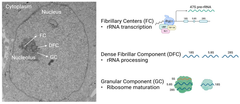
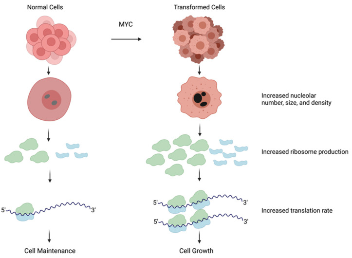
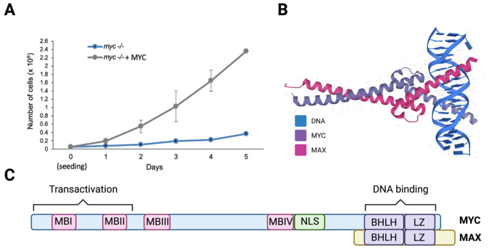
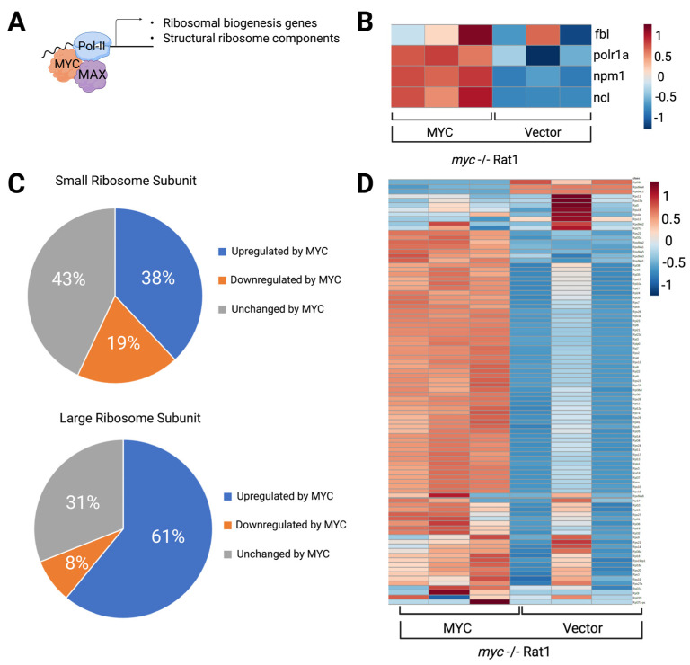
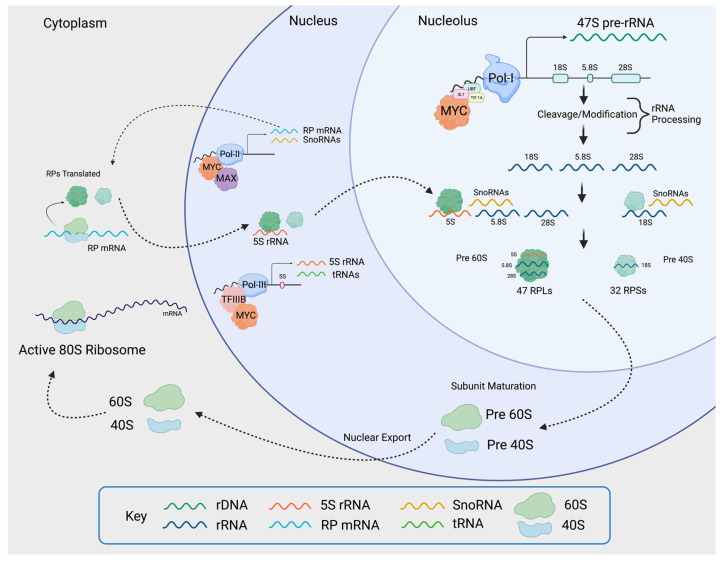
The nucleolus harbors the machinery necessary to produce new ribosomes which are critical for protein synthesis. Nucleolar size, shape, and density are highly dynamic and can be adjusted to accommodate ribosome biogenesis according to the needs for protein synthesis. In cancer, cells undergo continuous proliferation; therefore, nucleolar activity is elevated due to their high demand for protein synthesis. The transcription factor and universal oncogene MYC promotes nucleolar activity by enhancing the transcription of ribosomal DNA (rDNA) and ribosomal proteins. This review summarizes the importance of nucleolar activity in mammalian cells, MYC's role in nucleolar regulation in cancer, and discusses how a better understanding (and the potential inhibition) of aberrant nucleolar activity in cancer cells could lead to novel therapeutics.

Keywords: MYC; cell growth; nucleolus; ribosome; ribosome biogenesis; translation.


Conflict of interest statement

The authors declare no conflict of interest.

Figures

Figure 1
Figure 1
The nucleolus contains three distinct compartments responsible for pre-rRNA transcription, rRNA processing, and ribosome subunit assembly. The nucleoli are located within the nucleus (shown in the electron microscopy photo to the left) and are comprised of three sub-compartments: the fibrillary centers (FC), the dense fibrillar component (DFC), and the granular component (GC). The transcription of rDNA occurs in the FC upon the binding of selectivity factor 1 (SL1), which leads to the activation of the cofactors upstream binding factor (UBF) and TIF-1A, initiating RNAPolI to transcribe rDNA into 47S pre-rRNA. The 47S pre-rRNA is processed into 18S, 5.8S, and 28S rRNA in the DFC. Ribosome maturation proceeds in the GC, where additional RPs are wrapped around rRNAs. Figure created with Biorender.com (accessed on 31 January 2022).
Figure 2
Figure 2
Transformed cells have larger nucleoli and increased ribosome production. The nucleolus increases in size and density to accommodate the need for ribosome production. In transformed cells frequently due to hyperactivation of MYC, nucleoli are larger in size and darker in color, indicating higher activity, which results in an increase in ribosome number. Containing a larger number of ribosomes amplifies mRNA translation and ultimately leads to an increase in cell growth. Figure created with Biorender.com (accessed on 5 January 2022).
Figure 3
Figure 3
MYC heterodimerizes with MAX and increases cell proliferation. (A) Proliferation curve of HO15.19 myc-/- rat fibroblasts expressing empty vector or MYC. Cells were seeded and counted for 5 days. (B) The structure of the basic helix-loop-helix and leucine zipper (bHLH-LZ) domains of the heterodimer MYC-MAX and DNA (PDBe-KD), https://www.ebi.ac.uk/pdbe/pdbe-kb/proteins/P01106/interactions, (accessed on 28 November 2021). (C) Schematic representation of the MYC protein sequence with MYC Boxes (MBI, MBII, MBIII, MBIV), nuclear localization signal (NLS), and bHLHLZip domain on the C-terminus where DNA and MAX interacts. Figure created using Biorender.com (accessed on 28 January 2022).
Figure 4
Figure 4
Ribosomal biogenesis and structural ribosome components are upregulated in MYC-expressing cells. (A). Schematic of the interaction of MYC and MAX with the transcription machinery that drives the expression of regulatory and structural genes necessary for ribosome biogenesis. (B). Heatmap of myc-/- expressing empty vector or reconstituted with MYC. Data are extracted from published RNAseq [55] for nucleolar genes. (C). Pie chart showing that MYC increased the transcription of 38% of the structural components of the small ribosome subunit and 61% of the large subunit. Data were obtained by comparing the expression of structural ribosome genes in myc-/- expressing empty vector or reconstituted with MYC using a cutoff of Log2 fold change of or 0.58 and with adjusted p-value of 0.05. (D). Heatmap of myc-/- expressing empty vector or reconstituted by MYC. Data was extracted from published RNAseq [55] for structural RPs. Heatmaps were generated by MetaboAnalyst 5.0 (https://www.metaboanalyst.ca/, accessed on 10 December 2021). Figure created using Biorender.com, (accessed on 31 January 2022).
Figure 5
Figure 5
MYC induces ribosomal biogenesis processes. MYC heterodimerizes with MAX and promotes RNAPolI activity by binding to the rDNA promoter, as well as by activating the expression of selectivity factor 1 (SL1), which binds other RNAPolI cofactors such as upstream binding factor (UBF) and TIF-1A. The rDNA is transcribed into 47S pre-rRNA in the nucleolus. The pre-rRNA is processed and cleaved into 18S, 5.8S, and 28S rRNA. MYC-MAX simultaneously enhances RNAPolII activity by binding to RNAPolII-regulated promoters, as well as RNAPolIII activity by inducing the expression of the RNAPolIII cofactor TFIIIB. This yields RNAPolII-driven small RPs (RPS) mRNA, large RPs (RPL) mRNA, RNAPolIII-driven snoRNAs, and 5S rRNA. The mRNAs are transported into the cytoplasm where mature ribosomes translate them into small and large RPs. Once translated, small RPs are imported into the nucleolus. Large RPs are first imported into the nucleus, where they interact with 5S rRNA, and then to the nucleolus. In the nucleolus, the pre-rRNAs are modified and processed with the help of snoRNAs. As they are maturing, the rRNA wrap around the RPs, creating the pre-40 and pre-60 subunits which are exported to the cytoplasm for the final maturation step. Once maturation is complete in the cytoplasm, the active 80S ribosomes are formed. The small 40S subunit comprises 18S rRNA and 32 small RPs, and the large 60S subunit comprises 5S, 5.8S, and 28S rRNA and 47 large RPs. Figure created using Biorender.com (accessed on 31 January 2022).

References

    1. Montanaro L., Treré D., Derenzini M. Nucleolus, ribosomes, and cancer. Am. J. Pathol. 2008;173:301–310. doi: 10.2353/ajpath.2008.070752. - DOI - PMC - PubMed
    1. Derenzini M., Trerè D., Pession A., Montanaro L., Sirri V., Ochs R.L. Nucleolar function and size in cancer cells. Am. J. Pathol. 1998;152:1291–1297. - PMC - PubMed
    1. Thomas G. An encore for ribosome biogenesis in the control of cell proliferation. Nat. Cell Biol. 2000;2:E71–E72. doi: 10.1038/35010581. - DOI - PubMed
    1. Hernandez-Verdun D. Assembly and disassembly of the nucleolus during the cell cycle. Nucleus. 2011;2:189–194. doi: 10.4161/nucl.2.3.16246. - DOI - PMC - PubMed
    1. Henderson A.S., Warburton D., Atwood K.C. Location of ribosomal DNA in the human chromosome complement. Proc. Natl. Acad. Sci. USA. 1972;69:3394–3398. doi: 10.1073/pnas.69.11.3394. - DOI - PMC - PubMed

Publication types