. 2022 Jan 27;12(3):428.
doi: 10.3390/nano12030428.

Multimodal Contrast Agent Enabling pH Sensing Based on Organically Functionalized Gold Nanoshells with Mn-Zn Ferrite Cores

Affiliations

Multimodal Contrast Agent Enabling pH Sensing Based on Organically Functionalized Gold Nanoshells with Mn-Zn Ferrite Cores

Duong Thuy Bui et al. Nanomaterials (Basel). .

Abstract

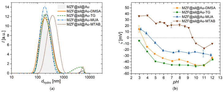
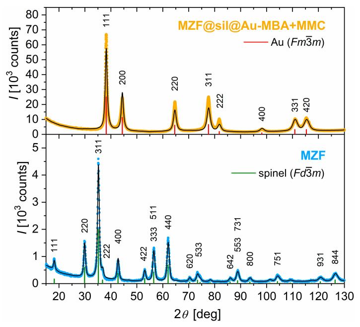
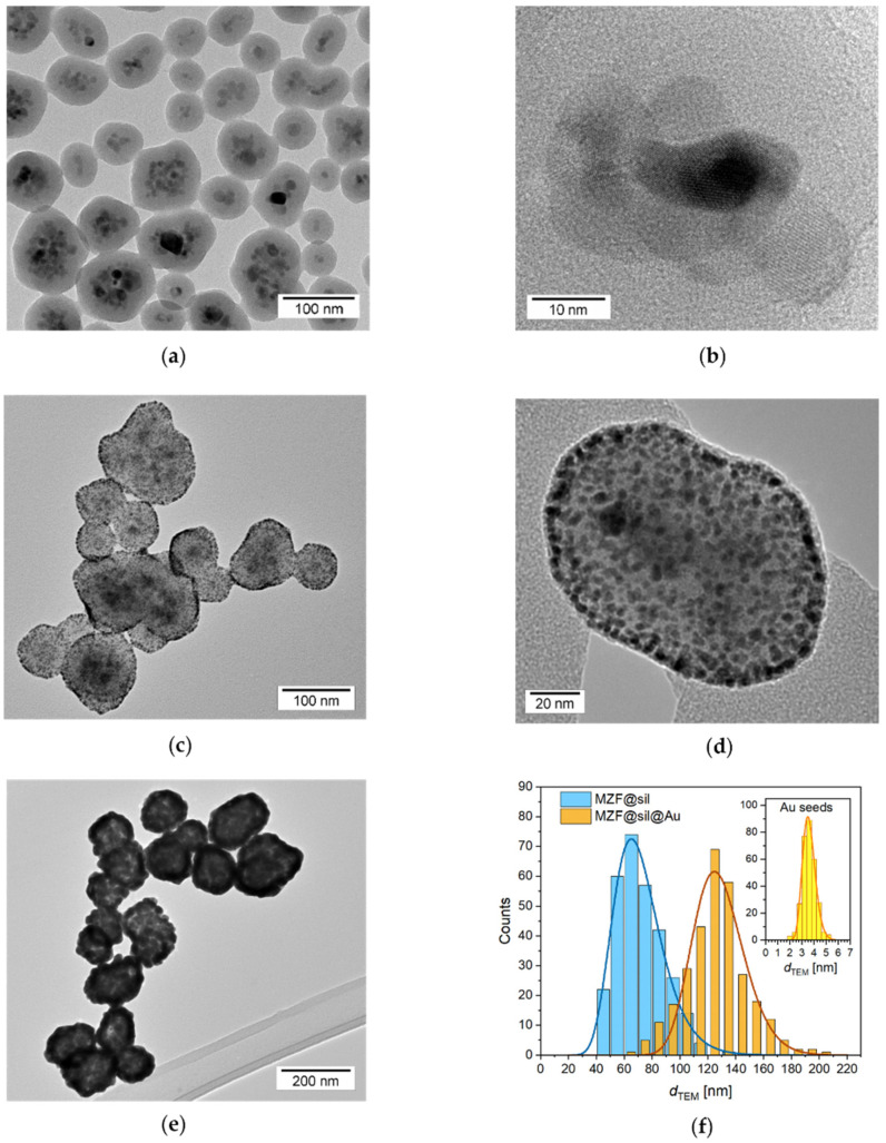
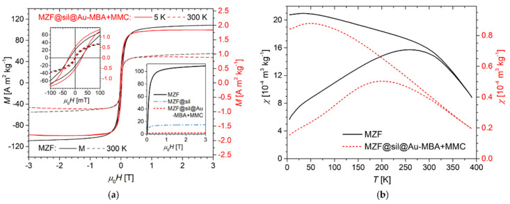
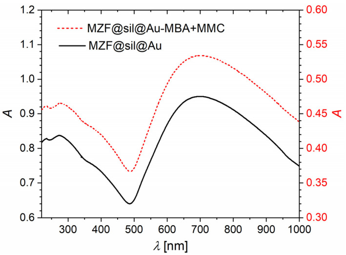
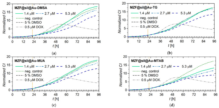
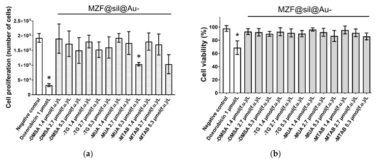
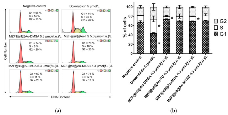
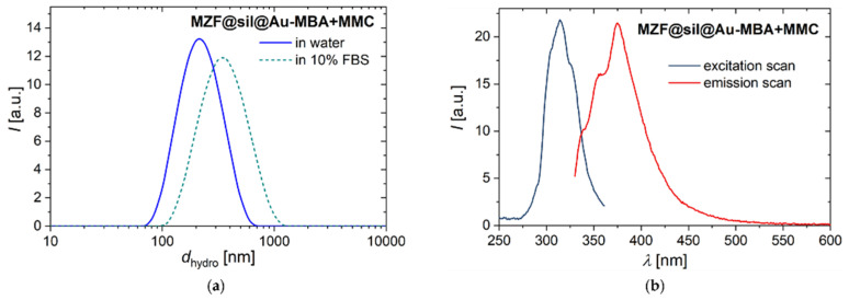
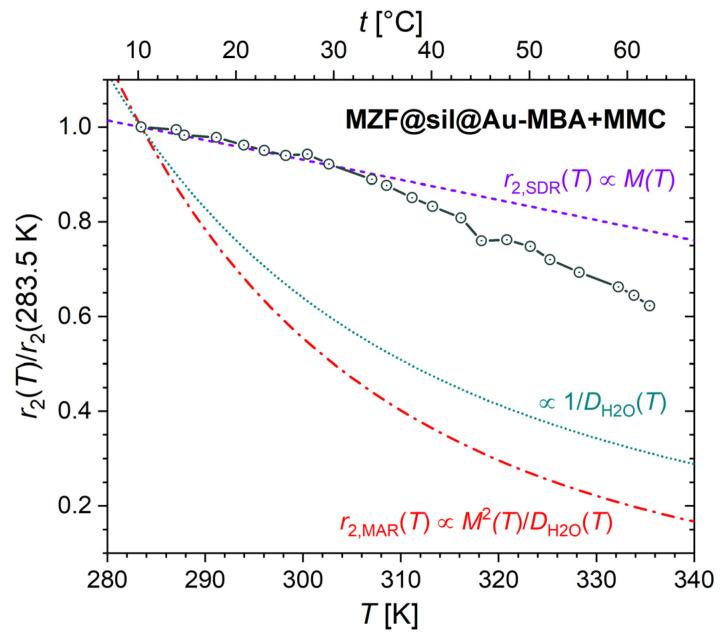
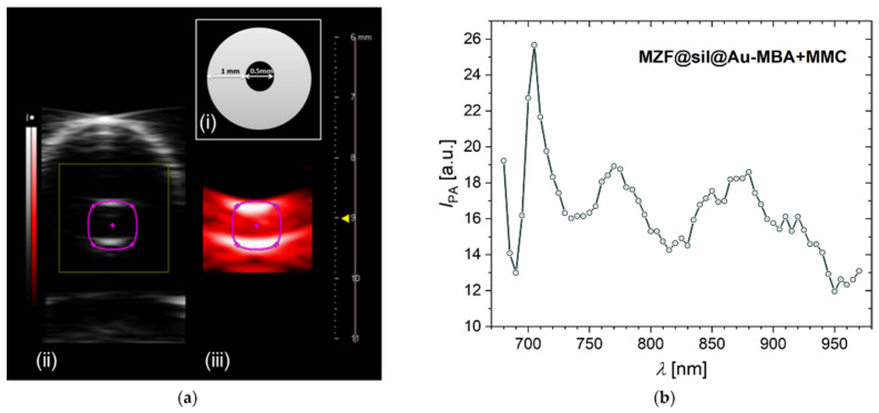
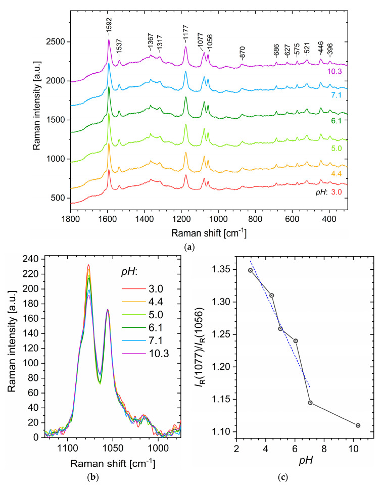
Highly complex nanoparticles combining multimodal imaging with the sensing of physical properties in biological systems can considerably enhance biomedical research, but reports demonstrating the performance of a single nanosized probe in several imaging modalities and its sensing potential at the same time are rather scarce. Gold nanoshells with magnetic cores and complex organic functionalization may offer an efficient multimodal platform for magnetic resonance imaging (MRI), photoacoustic imaging (PAI), and fluorescence techniques combined with pH sensing by means of surface-enhanced Raman spectroscopy (SERS). In the present study, the synthesis of gold nanoshells with Mn-Zn ferrite cores is described, and their structure, composition, and fundamental properties are analyzed by powder X-ray diffraction, X-ray fluorescence spectroscopy, transmission electron microscopy, magnetic measurements, and UV-Vis spectroscopy. The gold surface is functionalized with four different model molecules, namely thioglycerol, meso-2,3-dimercaptosuccinate, 11-mercaptoundecanoate, and (11-mercaptoundecyl)-N,N,N-trimethylammonium bromide, to analyze the effect of varying charge and surface chemistry on cells in vitro. After characterization by dynamic and electrophoretic light scattering measurements, it is found that the particles do not exhibit significant cytotoxic effects, irrespective of the surface functionalization. Finally, the gold nanoshells are functionalized with a combination of 4-mercaptobenzoic acid and 7-mercapto-4-methylcoumarin, which introduces a SERS active pH sensor and a covalently attached fluorescent tag at the same time. 1H NMR relaxometry, fluorescence spectroscopy, and PAI demonstrate the multimodal potential of the suggested probe, including extraordinarily high transverse relaxivity, while the SERS study evidences a pH-dependent spectral response.

Keywords: cell viability; gold nanoshells; magnetic nanoparticles; photoacoustic imaging; surface-enhanced Raman spectroscopy; transverse relaxivity.


Conflict of interest statement

The authors declare no conflict of interest.

Figures

Figure 5
Figure 5
Dynamic and electrophoretic light scattering studies on gold nanoshells functionalized with four different model molecules (DMSA, TG, MUA, MTAB): (a) The intensity-weighted hydrodynamic size distribution of gold nanoshells functionalized with the four model compounds, measured on dilute aqueous suspensions by DLS; (b) dependence of zeta potential on pH measured by ELS; the error bars show 95% confidence intervals based on repeated measurements.
Figure 1
Figure 1
Powder XRD patterns of the starting Mn0.61Zn0.42Fe1.97O4 nanoparticles (MZF) and the final organically functionalized gold nanoshells with silica-coated ferrite cores (MZF@sil@Au-MBA+MMC). The measured data are complemented with the calculated patterns (black lines) based on the Rietveld refinement of the spinel (Fd3¯m) and gold (Fm3¯m ) structures (original structure data retrieved from the Inorganic Crystal Structure Database under the collection codes 98553 and 64701). The diffraction lines of the refined structures are depicted by green and red verticals, respectively.
Figure 2
Figure 2
Transmission electron micrographs and size distribution of nanoparticles: (a) MZF@sil; (b) HRTEM detail of a magnetic core of MZF@sil particles; (c,d) MZF@sil nanoparticles decorated with gold seeds; (e) MZF@sil@Au; (f) histograms of MZF@sil and MZF@sil@Au sizes, with the inset showing the histogram of Au seeds on the surface of the decorated intermediate; the histograms were fitted with a log-normal distribution function.
Figure 3
Figure 3
Magnetic properties: (a) Hysteresis loops of bare ferrite nanoparticles (left axis) and final gold nanoshells (right axis) at temperatures of 5 K and 300 K (the scale of the right axis is adjusted so that the low-temperature curves of both samples coincide at μ0H = 0.3 T). The low-field details of magnetization curves are depicted in the upper left inset. The lower right inset shows the virgin curves for bare ferrite nanoparticles, silica-coated intermediate, and final gold nanoshells at 5 K. (b) ZFC-FC mass susceptibilities, χZFC and χFC, of bare ferrite nanoparticles (left axis) and gold nanoshells (right axis) in the magnetic field of 2 mT.
Figure 4
Figure 4
UV-Vis spectra of bare gold nanoshells (MZF@sil@Au, left axis) and gold nanoshells functionalized with the combination of MBA and MMC (MZF@sil@Au-MBA+MMC, right axis).
Figure 6
Figure 6
Growth kinetics of human MZF-7 breast carcinoma cells incubated with functionalized gold nanoshells as monitored by the xCELLigence system: Cells treated with (a) MZF@sil@Au-DMSA; (b) MZF@sil@Au-TG; (c) MZF@sil@Au-MUA; (d) MZF@sil@Au-MTAB of given concentrations. The negative (vehicle) control was treated with sterile deionized water, whereas the cells treated with 5% DMSO and 0.5 μM doxorubicin were used as positive controls. The yellow vertical line marks the time point of the treatment.
Figure 7
Figure 7
The effect of MZF@sil@Au-DMSA, MZF@sil@Au-TG, MZF@sil@Au-MUA, or MZF@sil@Au-MTAB on the (a) proliferation and (b) viability of MCF-7 cells. Changes in the proliferation and viability were monitored using the trypan blue exclusion test 48 h after the treatment. Results are shown as mean ± SD from three experiments; the asterisk marks results significantly different from the control (p ≤ 0.05). Cells treated with 1 µM doxorubicin and sterile deionized water were used as a positive and negative control, respectively.
Figure 8
Figure 8
Analysis of the cell cycle of MCF-7 cells 48 h after the application of suspensions of gold nanoshells functionalized with DMSA, TG, MUA, and MTAB with a ferrite concentration of 5.3 µmol(f.u.) L−1: (a) Representative histograms with the mean percentage of cells cycling through phases G1, S, and G2 from flow cytometry of three separate treatments; (b) the bar graph summarizing the percentage of cells in each phase of the cell cycle. Data are presented as mean values ± SD from three experiments; the asterisk marks the results significantly different (p ≤ 0.05) from the negative control that was treated with sterile deionized water. Cells treated with 5 µM doxorubicin were used as a positive control.
Figure 9
Figure 9
(a) The intensity-weighted hydrodynamic size distribution of MZF@sil@Au-MBA+MMC nanoshells in pure water and 10 vol% FBS measured by DLS. (b) Fluorescence spectra of MZF@sil@Au-MBA+MMC particles in an aqueous suspension with pH = 7. The excitation scan was recorded at λem = 375 nm, and the emission scan was measured with λex = 314 nm.
Figure 10
Figure 10
Temperature dependence of transverse relaxivity, r2, normalized by using the value at the lowest experimental temperature, r2(283.5 K), for MZF@sil@Au-MBA+MMC nanoshells in the magnetic field of 0.47 T. The dependence is compared with the dependences predicted for SDR (r2,SDR) and MAR (r2,MAR) and with the temperature dependence of the inverse value of the self-diffusion coefficient of water 1/DH2O(T).
Figure 11
Figure 11
Photoacoustic study on the suspension of MZF@sil@Au-MBA+MMC nanoshells with a ferrite concentration of 0.64 mmol(f.u.) L−1. (a) PAI study: (i) A scheme showing the experimental setup—a cross-section of the tube filled with the suspension, (ii) an ultrasound image of the tube with the nanoshells, and (iii) a photoacoustic image of the same tube obtained at 680-nm excitation; (b) photoacoustic spectrum of the MZF@sil@Au-MBA+MMC nanoshells.
Figure 12
Figure 12
SERS study on aqueous suspensions of MZF@sil@Au-MBA+MMC nanoparticles whose pH was adjusted in the range of 3–10 using diluted NaOH or HCl. (a) Raw (unprocessed) SERS spectra measured on aqueous suspensions with varying pH and (b) detail of the 1125–975 cm−1 region normalized to the MMC band at 1056 cm−1 with the pH-dependent intensity of the MBA band at 1077cm−1. (c) The dependence of the ratio of Raman band intensities at 1077 cm−1 and 1056 cm−1 on pH, complemented by linear fit in the range of pH = 3–7.

References

    1. Gleich B., Weizenecker R. Tomographic imaging using the nonlinear response of magnetic particles. Nature. 2005;435:1214–1217. doi: 10.1038/nature03808. - DOI - PubMed
    1. Lee N., Yoo D., Ling D., Cho M.H., Hyeon T., Cheon J. Iron oxide based nanoparticles for multimodal imaging and magnetoresponsive therapy. Chem. Rev. 2015;115:10637–10689. doi: 10.1021/acs.chemrev.5b00112. - DOI - PubMed
    1. Cui X.J., Mathe D., Kovacs N., Horvath I., Jauregui-Osoro M., de Rosales R.T.M., Mullen G.E.D., Wong W., Yan Y., Kruger D., et al. Synthesis, Characterization, and application of core shell Co0.16Fe2.84O4@NaYF4(Yb, Er) and Fe3O4@NaYF4(Yb, Tm) nanoparticle as trimodal (MRI, PET/SPECT, and Optical) imaging agents. Bioconjug. Chem. 2016;27:319–328. doi: 10.1021/acs.bioconjchem.5b00338. - DOI - PMC - PubMed
    1. Xing H.Y., Bu W.B., Zhang S.J., Zheng X.P., Li M., Chen F., He Q.J., Zhou L.P., Peng W.J., Hua Y.Q., et al. Mul-tifunctional nanoprobes for upconversion fluorescence, MR and CT trimodal imaging. Biomaterials. 2012;33:1079–1089. doi: 10.1016/j.biomaterials.2011.10.039. - DOI - PubMed
    1. Song G.S., Zheng X.C., Wang Y.J., Xia X., Chu S., Rao J.H. A Magneto-optical nanoplatform for multimodality imaging of tumors in mice. ACS Nano. 2019;13:7750–7758. doi: 10.1021/acsnano.9b01436. - DOI - PubMed

LinkOut - more resources