

Plumeria alba-Mediated Green Synthesis of Silver Nanoparticles Exhibits Antimicrobial Effect and Anti-Oncogenic Activity against Glioblastoma U118 MG Cancer Cell Line
- PMID: 35159838
- PMCID: PMC8839720
- DOI: 10.3390/nano12030493
Plumeria alba-Mediated Green Synthesis of Silver Nanoparticles Exhibits Antimicrobial Effect and Anti-Oncogenic Activity against Glioblastoma U118 MG Cancer Cell Line
Abstract
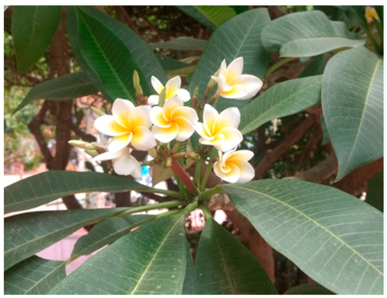
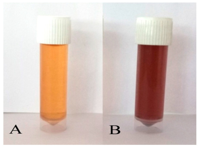
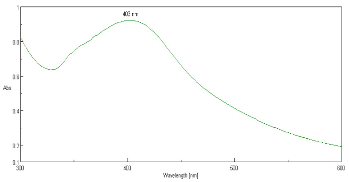
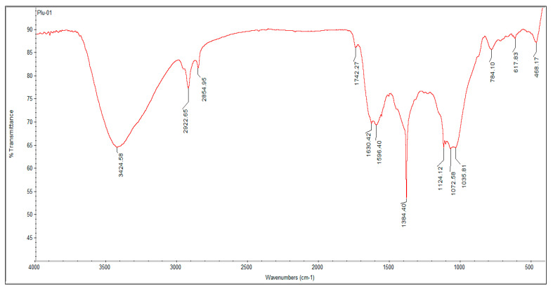
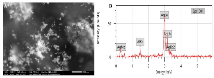
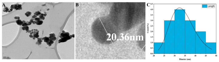
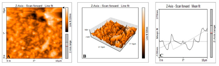
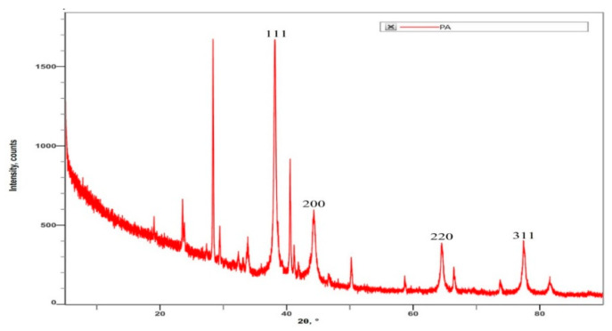
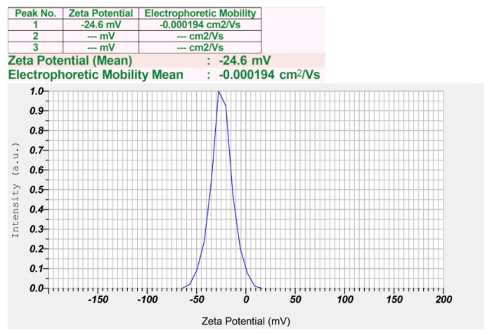
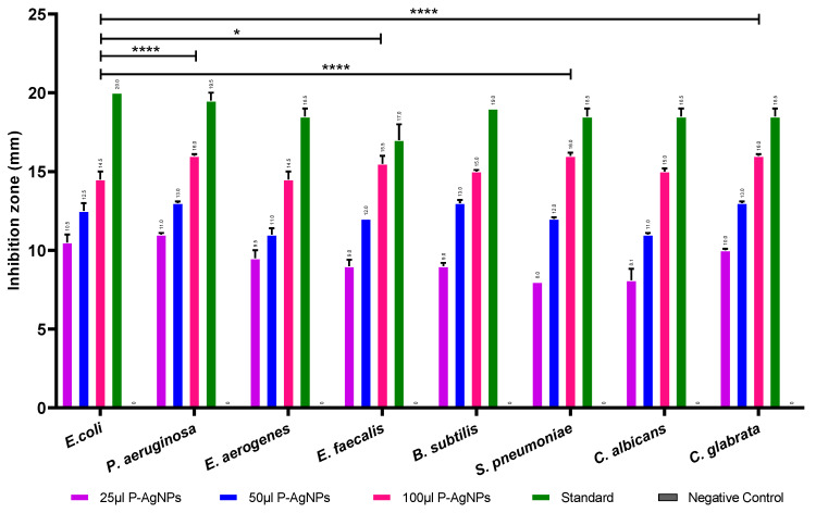
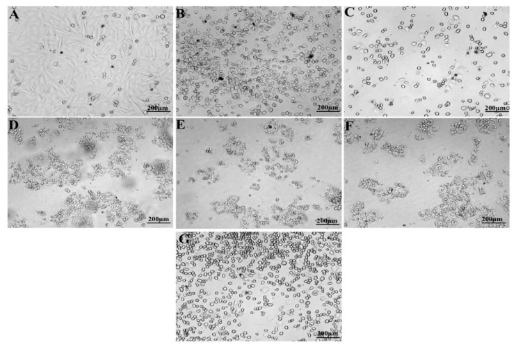
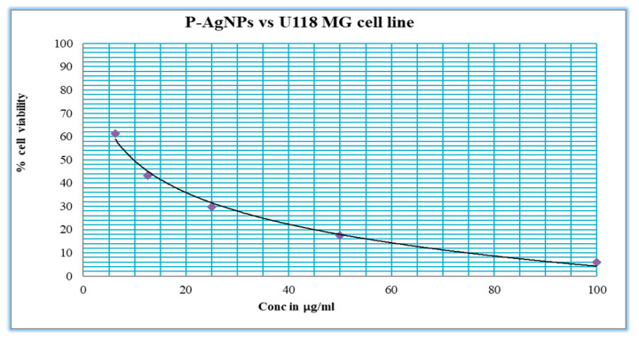
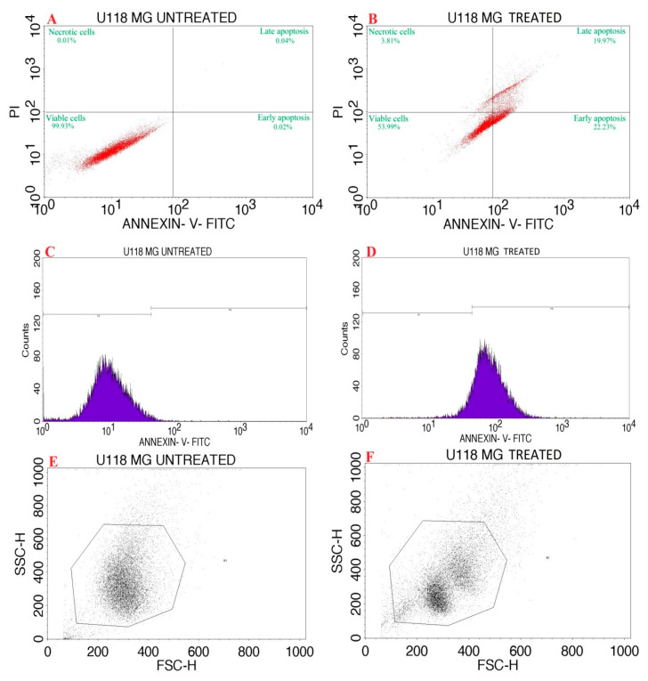
Plumeria alba (P. alba) is a small laticiferous tree with promising medicinal properties. Green synthesis of nanoparticles is eco-friendly, cost-effective, and non-hazardous compared to chemical and physical synthesis methods. Current research aiming to synthesize silver nanoparticles (AgNPs) from the leaf extract of P. alba (P- AgNPs) has described its physiochemical and pharmacological properties in recognition of its therapeutic potential as an anticancer and antimicrobial agent. These biogenic synthesized P-AgNPs were physiochemically characterized by ultraviolet-visible spectroscopy, Fourier-transform infrared spectroscopy (FTIR), scanning electron microscopy (SEM), transmission electron microscope (TEM), atomic force microscopy (AFM), X-ray diffractometry (XRD), and zeta potential analysis. Antimicrobial activity was investigated against Escherichia coli, Pseudomonas aeruginosa, Enterobacter aerogenes, Enterococcus faecalis, Bacillus subtilis, Streptococcus pneumoniae, Candida albicans, and Candida glabrata. Anticancer activity against glioblastoma U118 MG cancer lines was investigated using an MTT assay, and apoptosis activity was determined by flow cytometry. UV-visible spectroscopic analysis portrayed surface plasmon resonance at 403 nm of synthesized P-AgNPs, and FTIR suggested the presence of amines, alkanes, and phenol molecules that could be involved in reduction and capping processes during AgNPs formation. Synthesized particles were spherical in shape and poly-dispersed with an average particle size of 26.43 nm and a poly-dispersity index (PDI) of 0.25 with a zeta potential value of -24.6 mV, ensuring their stability. The lattice plane values confirm the crystalline nature as identified by XRD. These P-AgNPs exhibited potential antimicrobial activity against selected human pathogenic microbes. Additionally, the in vitro MTT assay results showed its effective anticancer activity against the glioma U118 MG cancer cell line with an IC50 value of 9.77 µg/mL AgNPs by initiating apoptosis as identified by a staining study with flow cytometric Annexin V-Fluorescein Isothiocyanate (FITC) and Propidium Iodide (PI). Thus, P. alba AgNPs can be recommended for further pharmacological and other biological research. To conclude, the current investigation developed an eco-friendly AgNPs synthesis using P. alba leaf extract with potential cytotoxic and antibacterial capacity, which can therefore be recommended as a new strategy to treat different human diseases.
Keywords: Apocynaceae; Plumeria alba; apoptosis; chemotherapeutics; cytotoxic; nano-antibiotics; nanotechnology.
Conflict of interest statement
The authors declare that there is no conflict of interest.
Figures
References
-
- Moodley J.S., Babu Naidu Krishna S., Pillay K., Sershen, Govender P. Green synthesis of silver nanoparticles from Moringa oleifera leaf extracts and its antimicrobial potential. Adv. Nat. Sci. Nanosci. Nanotechnol. 2018;9:015011. doi: 10.1088/2043-6254/aaabb2. - DOI
-
- Khan I., Saeed K., Khan I. Nanoparticles: Properties, applications and toxicities. Arab. J. Chem. 2019;12:908–931. doi: 10.1016/j.arabjc.2017.05.011. - DOI
Grants and funding
LinkOut - more resources
Full Text Sources
Research Materials
Miscellaneous
