

Comparison between Two Different Scanners for the Evaluation of the Role of 18F-FDG PET/CT Semiquantitative Parameters and Radiomics Features in the Prediction of Final Diagnosis of Thyroid Incidentalomas
- PMID: 35160067
- PMCID: PMC8836668
- DOI: 10.3390/jcm11030615
Comparison between Two Different Scanners for the Evaluation of the Role of 18F-FDG PET/CT Semiquantitative Parameters and Radiomics Features in the Prediction of Final Diagnosis of Thyroid Incidentalomas
Abstract
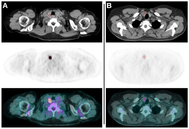
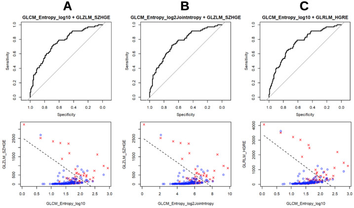
The aim of this study was to compare two different tomographs for the evaluation of the role of semiquantitative PET/CT parameters and radiomics features (RF) in the prediction of thyroid incidentalomas (TIs) at 18F-FDG imaging. A total of 221 patients with the presence of TIs were retrospectively included. After volumetric segmentation of each TI, semiquantitative parameters and RF were extracted. All of the features were tested for significant differences between the two PET scanners. The performances of all of the features in predicting the nature of TIs were analyzed by testing three classes of final logistic regression predictive models, one for each tomograph and one with both scanners together. Some RF resulted significantly different between the two scanners. PET/CT semiquantitative parameters were not able to predict the final diagnosis of TIs while GLCM-related RF (in particular GLCM entropy_log2 e GLCM entropy_log10) together with some GLRLM-related and GLZLM-related features presented the best predictive performances. In particular, GLCM entropy_log2, GLCM entropy_log10, GLZLM SZHGE, GLRLM HGRE and GLRLM HGZE resulted the RF with best performances. Our study enabled the selection of some RF able to predict the final nature of TIs discovered at 18F-FDG PET/CT imaging. Classic semiquantitative and volumetric PET/CT parameters did not reveal these abilities. Furthermore, a good overlap in the extraction of RF between the two scanners was underlined.
Keywords: 18F-FDG; PET/CT; positron emission tomography; radiomics; texture analysis; thyroid cancer; thyroid incidentalomas.
Conflict of interest statement
The authors declare no conflict of interest.
Figures

References
-
- Stokkel M.P., Duchateau C.S., Dragoiescu C. The value of FDG-PET in the follow-up of differentiated thyroid cancer: A review of the literature. Q. J. Nucl. Med. Mol. Imaging. 2006;50:78–87. - PubMed
LinkOut - more resources
Full Text Sources
