. 2022 Jan 30;11(3):760.
doi: 10.3390/jcm11030760.

Ankle Magnetic Resonance Imaging in Juvenile Idiopathic Arthritis Versus Non-Juvenile Idiopathic Arthritis Patients with Arthralgia

Affiliations

Ankle Magnetic Resonance Imaging in Juvenile Idiopathic Arthritis Versus Non-Juvenile Idiopathic Arthritis Patients with Arthralgia

Monika Ostrowska et al. J Clin Med. .

Abstract

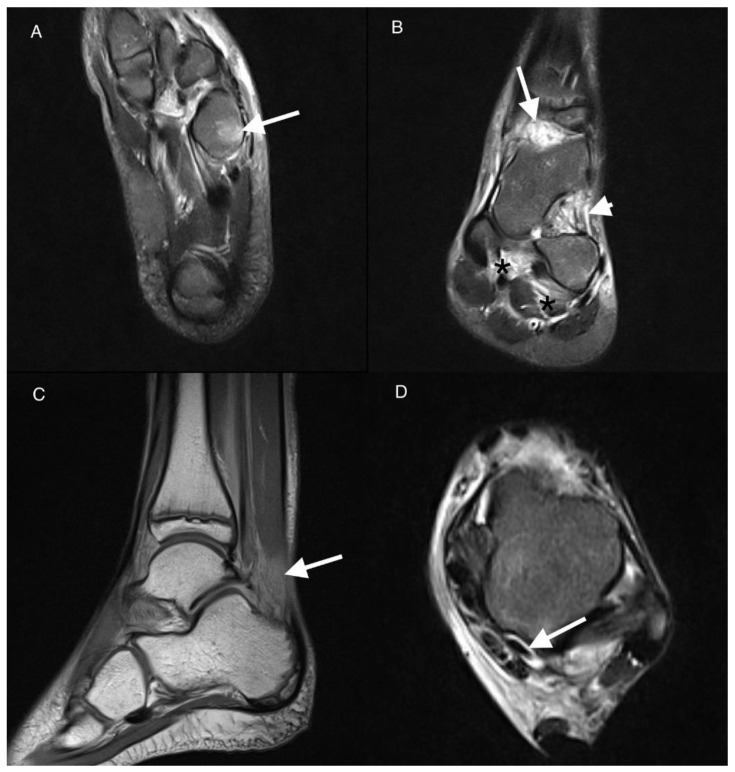
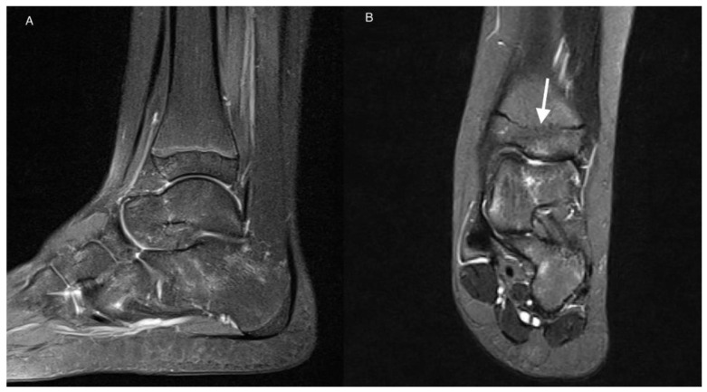
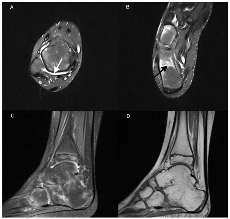
This retrospective case-control study aimed to evaluate whether Magnetic Resonance Imaging (MRI) enables differentiation of ankle arthritis in Juvenile Idiopathic Afrthritis JIA from ankle arthralgia of unknown aetiology in patients clinically suspected of JIA. Forty-four children, at ages 5-16, who underwent MRI of the ankle from January 2016 to March 2021 for clinically suspected active ankle arthritis in the course of JIA were included. MRI findings in both groups-patients with the final diagnosis of JIA and without final diagnosis of JIA-were compared and scored. The sum of the scores of 22 ankle lesions in an individual patient (active, destructive and developmental), so-called the MRI summarized score, was calculated and tested in terms of the most optimal diagnosis of JIA. Interobserver agreement was calculated. Inflammatory features were seen on MRI in 38 out of all the included patients (86%). The most common lesions in both groups were effusion in the tibio-talar joint (68% in JIA and 64% in the arthralgia group) and effusion in subtalar joint (64% in JIA vs. 59% in the arthralgia group). In general, more lesions were identified in the JIA group than in non-JIA. However, only tenosynovitis was significantly more common in the JIA vs. non-JIA group (p = 0.031). The MRI summarized score did not allow for discrimination between ankle arthritis in JIA from non-JIA patients; the best levels of sensitivity (32%), specificity (91%), positive predictive value PPV (78%) and negative predictive value NPV (57%) were achieved only at the cut-off point of 10.

Keywords: ankle arthritis; juvenile idiopathic arthritis; magnetic resonance imaging; scoring.


Conflict of interest statement

The authors declare no conflict of interest.

Figures

Figure 1
Figure 1
Magnetic resonance imaging (MRI) of the left ankle of an 11-year-old boy with juvenile idiopathic arthritis (JIA). T2-weighted turbo invertion recovery magnitude (TIRM) images in axial (A,D), coronal (B), and sagittal PD-weighted planes (C). Bone marrow edema (BME) stage 2 in the cuboid bone (arrow in (A)). Effusion/synovial thickening stage 2 in the tibio-talar joint (arrow in (B)). Fat tissue in sinus tarsi involvement stage 1 (arrowhead in (B)). Enthesitis of the interosseous ligaments stage 1 (black asterisks in (B)). Kager’s fat pad inflammation arrow in (C). Tenosynovitis of the flexor hallucis longus muscle tendon stage 1 arrow in (D).
Figure 2
Figure 2
MRI of the left ankle of an 11-year-old girl with JIA. T2-weighted TIRM images in sagittal (A) and coronal (B) planes. Patchy BME (stage 1) in talus and calcaneus. Developmental disorder in the form of premature closure of physis (arrow).
Figure 3
Figure 3
MRI of the right ankle of a 9-year-old boy with JIA. Axial T2-weighted TIRM images (A,B), sagittal PD with fat saturation (C), and sagittal PD-weighted image (D). Effusion/synovial thickening stage 2 in the tibio-talar joint (arrow). BME in numerous bones of the ankle and tarsum, the most advanced in calcaneus stage 3 (arrow in (B)). Tibio-talar joint space narrowing, tibio-calcaneal ankylosis, and osteophytes (C,D).

Similar articles

Cited by

References

    1. Hemke R., Kuijpers T.W., van der Berg J.M., Van Veenendaal M., Dolman K.M., Van Rossum M.A.J., Maas M. The diagnostic accuracy of unenhanced MRI in the assessment of joint abnormalities in juvenile idiopathic arthritis. Eur. Radiol. 2013;23:1998–2004. doi: 10.1007/s00330-013-2770-9. - DOI - PubMed
    1. Raveli A., Martini A. Juvenile idiopathic arthritis. Lancet. 2007;369:767–778. doi: 10.1016/S0140-6736(07)60363-8. - DOI - PubMed
    1. Pascoli L., Wright S., McAllister C., Rooney M. Prospective evaluation of clinical and ultrasound findings in ankle disease in juvenile idiopathic arthritis: Importance of ankle ultrasound. J. Rheumatol. 2010;37:2409–2414. doi: 10.3899/jrheum.091262. - DOI - PubMed
    1. Peters S.E., Laxer R.M., Connolly B.L., Parra D.A. Ultrasound—guided steroid tendon sheath injection in juvenile idiopathic arthritis: A 10-year single center retrospective study. Pediatr. Rheumatol. 2017;15:22–29. doi: 10.1186/s12969-017-0155-3. - DOI - PMC - PubMed
    1. Lovell D.J. Juvenile idiopathic arthritis: Clinical features. In: Klippel J.H., Stone J.H., Crofford L.J., White P.F., editors. Primer on the Rheumatic Diseases. Springer; New York, NY, USA: 2008. pp. 142–148.

LinkOut - more resources