

Usefulness of Dermoscopy in Localized Scleroderma (LoS, Morphea) Diagnosis and Assessment-Monocentric Cross-Sectional Study
- PMID: 35160216
- PMCID: PMC8836985
- DOI: 10.3390/jcm11030764
Usefulness of Dermoscopy in Localized Scleroderma (LoS, Morphea) Diagnosis and Assessment-Monocentric Cross-Sectional Study
Abstract
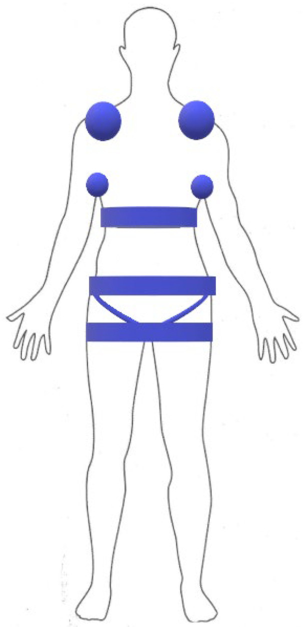
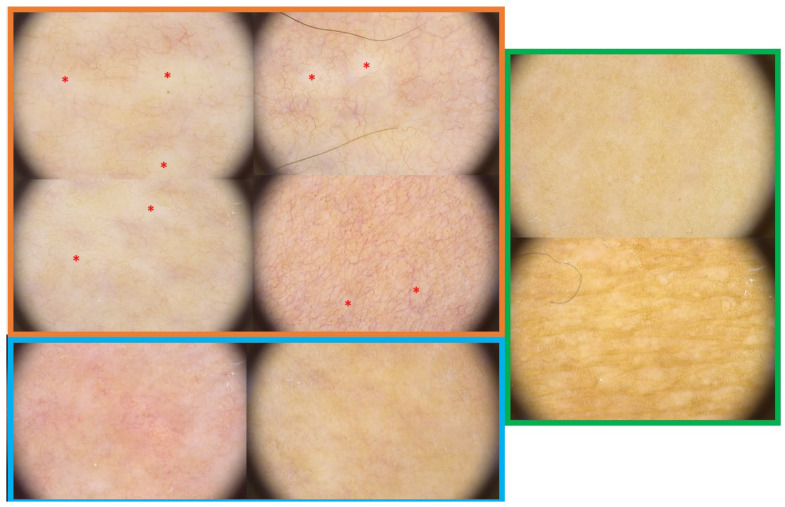
Morphea, also known as localized scleroderma (LoS), is a chronic autoimmune disease of the connective tissue. The clinical picture of LoS distinguishes between active and inactive lesions. Sometimes the clinical findings are challenging to identify, and therefore, the need for additional methods is emphasized. Our study aimed to demonstrate the characteristic dermoscopic features in morphea skin lesions, focusing on demonstrating features in active and inactive lesions. In our patients (n = 31) with histopathologically proven LoS, we performed clinical evaluation of lesions (n = 162): active/inactive and according to both disease activity (modified localized scleroderma severity index, mLoSSI) and damage (localized scleroderma skin damage index, LoSDI) parameters. In addition, we took into account compression locations to determine whether skin trauma, a known etiopathogenetic factor in LoS, affects the dermoscopic pattern of the lesions. We performed a dermoscopy of the lesions, categorizing the images according to the severity within the observed field. We showed that within the active lesions (clinically and with high mLoSSI), white clouds and linear branching vessels had the highest severity. These features decreased within the observed field in inactive lesions and with high LoSDI. Brownish structureless areas were most intense in inactive lesions with high LoSDI. Erythematous areas, linear branching vessels, dotted vessels, and crystalline structures were statistically significant for pressure locations. We have shown dermoscopy is a valuable tool to assess the activity or inactivity of lesions, which translates into appropriate therapeutic decisions and the possibility of monitoring the patient during and after therapy for possible relapse.
Keywords: dermoscopy; localized scleroderma; morphea.
Conflict of interest statement
The authors declare no conflict of interest.
Figures
References
-
- Krasowska D., Rudnicka L., Dańczak-Pazdrowska A., Chodorowska G., Woźniacka A., Lis-Święty A., Czuwara J., Maj J., Majewski S., Sysa-Jędrzejowska A., et al. Localized scleroderma (morphea). Diagnostic and therapeutic recommendations of the Polish Dermatological Society. Dermatol. Rev. 2019;106:333–353. doi: 10.5114/dr.2019.88252. - DOI
-
- Errichetti E., Stinco G. Morphea. In: Micali G., Lacarrubba F., Stinco G., Argenziano G., Neri I., editors. Atlas of Pediatric Dermatoscopy. Springer; Cham, Switzerland: 2018. pp. 115–119. - DOI
-
- Knobler R., Moinzadeh P., Hunzelmann N., Kreuter A., Cozzio A., Mouthon L., Cutolo M., Rongioletti F., Denton C., Rudnicka L., et al. European Dermatology Forum S1-guideline on the diagnosis and treatment of sclerosing diseases of the skin, Part 1: Localized scleroderma, systemic sclerosis and overlap syndromes. J. Eur. Acad. Dermatol. Venereol. 2017;31:1401–1424. doi: 10.1111/jdv.14458. - DOI - PubMed
LinkOut - more resources
Full Text Sources
