Review
. 2022 Feb 7;11(3):860.
doi: 10.3390/jcm11030860.

Viscoelastic Hemostatic Assays: A Primer on Legacy and New Generation Devices

Affiliations
Review

Viscoelastic Hemostatic Assays: A Primer on Legacy and New Generation Devices

Oksana Volod et al. J Clin Med. .

Abstract

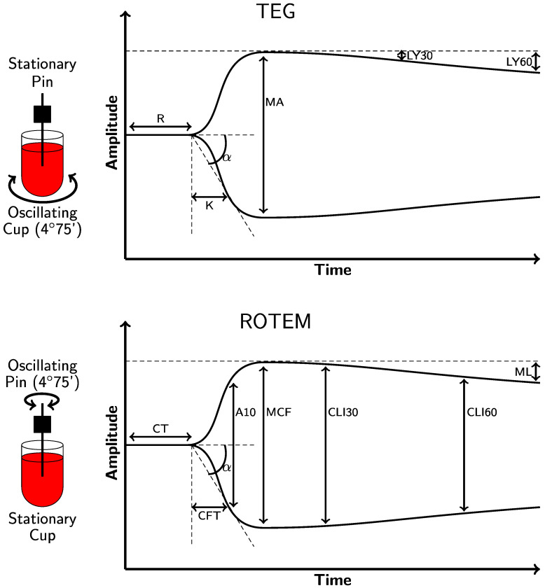
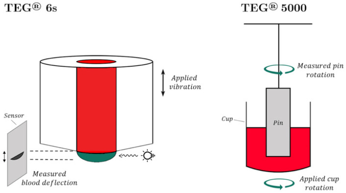
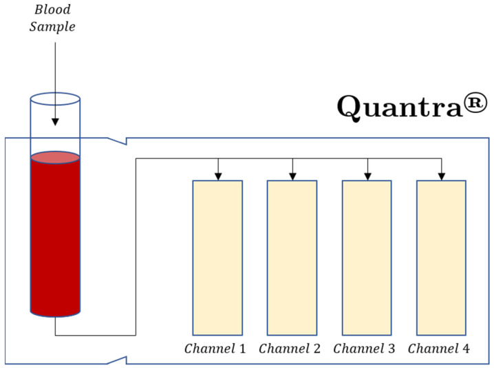
Viscoelastic hemostatic assay (VHAs) are whole blood point-of-care tests that have become an essential method for assaying hemostatic competence in liver transplantation, cardiac surgery, and most recently, trauma surgery involving hemorrhagic shock. It has taken more than three-quarters of a century of research and clinical application for this technology to become mainstream in these three clinical areas. Within the last decade, the cup and pin legacy devices, such as thromboelastography (TEG® 5000) and rotational thromboelastometry (ROTEM® delta), have been supplanted not only by cartridge systems (TEG® 6S and ROTEM® sigma), but also by more portable point-of-care bedside testing iterations of these legacy devices (e.g., Sonoclot®, Quantra®, and ClotPro®). Here, the legacy and new generation VHAs are compared on the basis of their unique hemostatic parameters that define contributions of coagulation factors, fibrinogen/fibrin, platelets, and clot lysis as related to the lifespan of a clot. In conclusion, we offer a brief discussion on the meteoric adoption of VHAs across the medical and surgical specialties to address COVID-19-associated coagulopathy.

Keywords: COVID-19; coagulopathy; fibrinogen; hemorrhage; heparin; personalized medicine; rotational thromboelastometry; thromboelastography; thrombosis.


Conflict of interest statement

O.V. is a consultant for Haemonetics Corporation (Boston, MA, USA), served on the Clinical Care Improvement Steering Committee for Diagnostica Stago, Inc., and received honoraria for consultancy outside the submitted work. E.E.M., H.B.M., M.D.N. and M.M.W. have received research grants from Haemonetics Corporation outside the submitted work. M.D.N. has received an honorarium from Haemonetics Corporation for speaking engagements, as well as research support from Janssen Pharmaceuticals (Beerse, Belgium) and Noveome Biotherapeutics (Pittsburgh, PA, USA) outside the submitted work. He has served as a consultant to Janssen and CSL Behring (King of Prussia, PA, USA) and serves on the Scientific Advisory Board of Haima Therapeutics (Cleveland, OH, USA). M.M.W. has received honoraria from Alexion Pharmaceuticals (Boston, MA, USA). D.F. has received study funding, honoraria for consultancy, and shows board activity from Astra Zeneca, AOP orphan, Baxter, Bayer, B. Braun Medical, Biotest, CSL Behring, Delta Select, Dade Behring, Edwards, Fresenius, Glaxo, Haemoscope, Hemogem, Lilly, LFB, Mitsubishi Pharma, Novo Nordisk, Octapharma, Pfizer, and Tem Innovations outside the submitted work.

Figures

Figure 1
Figure 1
Depictions of the physiologic TEG® 5000 (top) and ROTEM® delta (bottom) tracings. As the left side of the figure illustrates, a pin descends into a cup containing a sample of whole blood that is maintained at 37 °C. TEG® 5000 and ROTEM® delta analyzers use equivalent parameters that are labeled differently. Reaction time (R) and clotting time (CT) measure the time required for the transducer to displace by 2 mm on the y-axis. Clot formation/kinetics (K) and clot formation time (CFT) measure the initial clot strength and the time needed to displace the transducer by 20 mm, measured from when it first reached 2 mm. The α-angle measures the rate of clot formation in both the TEG® 5000 and ROTEM® delta analyzers by analyzing the angle formed between the end of the R/CT (which is called the split point) and the 20 mm point on the y-axis. The fibrinogen level is broadly correlated with both the K/CFT and α-angle. The reference ranges and definitions of each parameter are provided in Table 1. The clot amplitude at 5 and 10 min (A5 and A10) measures the amplitude at 5 min intervals after the end of R/CT. The maximum amplitude (MA) and maximum clot firmness (MCF) measure the maximum displacement and are indicative of the maximum clot strength. They also correlate with the maximum clot retraction and reflect the crosslinking of fibrin with platelets. Fibrinolysis is depicted by differing parameters in the TEG® 5000 and ROTEM® delta analyzers. The lysis at 30 and 60 min (LY30 and LY60) is a measure of the percent of decrease in amplitude at 30 and 60 min after achieving MA. The clot lysis index (CLI30 and CLI60) is the residual clot remaining 30 and 60 min after CT measured as a percentage of MCF. The maximum lysis (ML) is a measure of the percent of decrease in amplitude at the end of the run [20,23,34,35,36,37,38,39,40,41].
Figure 2
Figure 2
(Left): TEG® 6s. The vibration of the sample induces blood deflection; the measures change with the resonance frequency. (Right): TEG® 5000. The difference in the rotations of the cup and pin is measured via a torsional spring [64,65].
Figure 3
Figure 3
The Quantra® plus cartridge has four channels with reagents designed to measure clot stiffness (CS) and clot time (CT). Channel 1 measures CT with kaolin activation. Channel 2 measures CT with kaolin activation and heparin neutralization. Channel 3 measures CS and tissue factor activation with heparin neutralization. Channel 4 measures CS with tissue factor activation, platelet inhibition, and heparin neutralization. Channel 4 measures the fibrinogen contribution to CS, (FCS). The platelet contribution to CS, (PCS) is equal to CS-FCS. Clot Time Ratio (CTR) = CT/CTH and indicates the level of heparinization of the patient blood sample [69,78].

References

    1. Starzl T.E. The saga of liver replacement, with particular reference to the reciprocal influence of liver and kidney transplantation (1955–1967) J. Am. Coll. Surg. 2002;195:587–610. doi: 10.1016/S1072-7515(02)01498-9. - DOI - PMC - PubMed
    1. Groth C.G., Pechet L., Starzl T.E. Coagulation during and after orthotopic transplantation of the human liver. Arch. Surg. 1969;98:31–34. doi: 10.1001/archsurg.1969.01340070049006. - DOI - PMC - PubMed
    1. Kang Y.G., Martin D.J., Marquez J., Lewis J.H., Bontempo F.A., Shaw B.W., Jr., Starzl T.E., Winter P.M. Intraoperative changes in blood coagulation and thrombelastographic monitoring in liver transplantation. Anesth. Analg. 1985;64:888–896. doi: 10.1213/00000539-198509000-00008. - DOI - PMC - PubMed
    1. Starzl T.E., Marchioro T.L., Vonkaulla K.N., Hermann G., Brittain R.S., Waddell W.R. Homotransplantation of the Liver in Humans. Surg. Gynecol. Obstet. 1963;117:659–676. - PMC - PubMed
    1. Shore-Lesserson L., Manspeizer H.E., DePerio M., Francis S., Vela-Cantos F., Ergin M.A. Thromboelastography-guided transfusion algorithm reduces transfusions in complex cardiac surgery. Anesth. Analg. 1999;88:312–319. doi: 10.1213/00000539-199902000-00016. - DOI - PubMed

LinkOut - more resources