. 2022 Jan 20;14(3):397.
doi: 10.3390/polym14030397.

Optimal Morphometric Characteristics of a Tubular Polymeric Scaffold to Promote Peripheral Nerve Regeneration: A Scoping Review

Affiliations

Optimal Morphometric Characteristics of a Tubular Polymeric Scaffold to Promote Peripheral Nerve Regeneration: A Scoping Review

Josefa Alarcón Apablaza et al. Polymers (Basel). .

Abstract

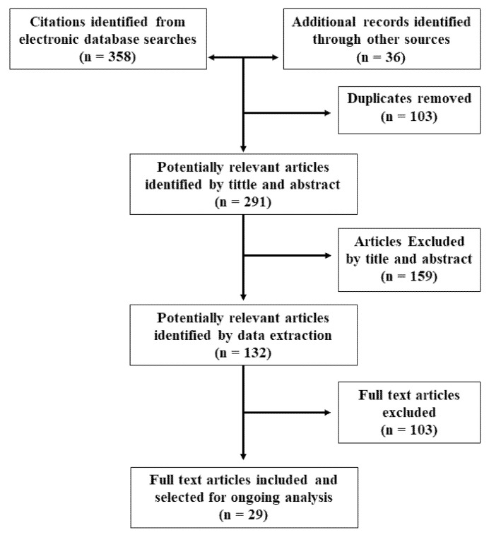
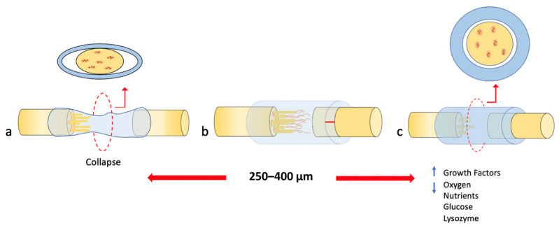
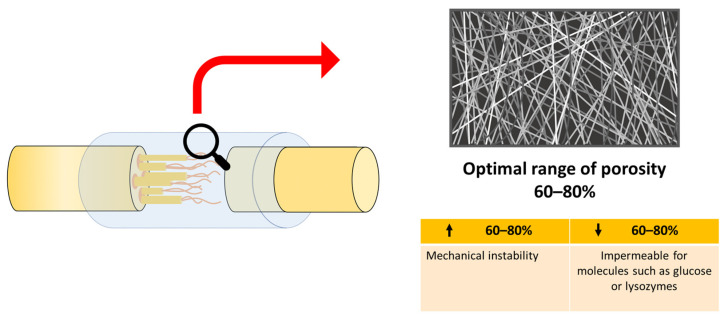
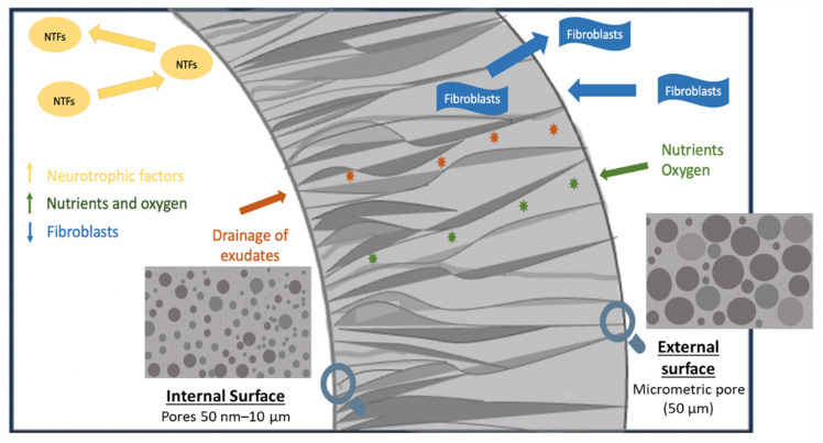
Cellular behavior in nerve regeneration is affected by the architecture of the polymeric nerve guide conduits (NGCs); therefore, design features of polymeric NGCs are critical for neural tissue engineering. Hence, the purpose of this scoping review is to summarize the adequate quantitative/morphometric parameters of the characteristics of NGC that provide a supportive environment for nerve regeneration, enhancing the understanding of a previous study. 394 studies were found, of which 29 studies were selected. The selected studies revealed four morphometric characteristics for promoting nerve regeneration: wall thickness, fiber size, pore size, and porosity. An NGC with a wall thickness between 250-400 μm and porosity of 60-80%, with a small pore on the inner surface and a large pore on the outer surface, significantly favored nerve regeneration; resulting in an increase in nutrient permeability, retention of neurotrophic factors, and optimal mechanical properties. On the other hand, the superiority of electrospun fibers is described; however, the size of the fiber is controversial in the literature, obtaining optimal results in the range of 300 nm to 30 µm. The incorporation of these optimal morphometric characteristics will encourage nerve regeneration and help reduce the number of experimental studies as it will provide the initial morphometric parameters for the preparation of an NGC.

Keywords: morphology; nerve scaffold; peripheral nerve regeneration; regenerative biology; tissue engineering.


Conflict of interest statement

All authors declare no conflict of interest. The funders had no role in the design of the study, in the collection, analyses, or interpretation of data, in the writing of the manuscript, or in the decision to publish the results.

Figures

Figure 1
Figure 1
Flow chart for study selection.
Figure 2
Figure 2
Extracted and modified from Part I of this study [24]. The behavior of the different wall thicknesses in the collapse of the NGC. (a) Shows a wall thickness lower than 250–400 μm resulting in the collapse of the NGC. (b) Shows the optimal wall thickness in a range of 250–400 μm resulting in successful nerve regeneration. (c) Shows a wall thickness greater than 250–400 μm resulting in greater retention of growth factors within the lumen, but decreased oxygen for and exchange of nutrients such as glucose and lysozyme through the walls.
Figure 3
Figure 3
Extracted and modified from Part I of this study [24]. Schematic diagram of optimal porosity. Lower porosity ( 60–80%) provides impermeable for molecules. Higher porosity ( 60–80%)) provides mechanically instability.
Figure 4
Figure 4
Graphic summary of the asymmetric NGC with pores 50 nm–10 μm on the inner surface and pores of 50 μm on the outer surface. This distribution of pore sizes improved nerve regeneration by minimizing the infiltration of fibrous tissue and the escape of neurotrophic factors (NTFs). In addition, asymmetric NGC facilitated the rapid drainage of exudates (orange stars) from nerve wounds, while providing high permeability of nutrients and oxygen (green stars).

References

    1. Madduri S., Gander B. Growth factor delivery systems and repair strategies for damaged peripheral nerves. J. Control. Release. 2012;161:274–282. doi: 10.1016/j.jconrel.2011.11.036. - DOI - PubMed
    1. Li R., Liu Z., Pan Y., Chen L., Zhang Z., Lu L. Peripheral nerve injuries treatment: A systematic review. Cell Biochem. Biophys. 2014;68:449–454. doi: 10.1007/s12013-013-9742-1. - DOI - PubMed
    1. Du J., Chen H., Qing L., Yang X., Jia X. Biomimetic neural scaffolds: A crucial step towards optimal peripheral nerve regeneration. Biomater. Sci. 2018;6:1299–1311. doi: 10.1039/C8BM00260F. - DOI - PMC - PubMed
    1. Kim J.I., Hwang T.I., Aguilar L.E., Park C.H., Kim C.S. Controlled Design of Aligned and Random Nanofibers for 3D Bi-functionalized Nerve Conduits Fabricated via a Novel Electrospinning Set-up. Sci. Rep. 2016;6:23761. doi: 10.1038/srep23761. - DOI - PMC - PubMed
    1. Jiang X., Mi R., Hoke A., Chew S.Y. Nanofibrous nerve conduit-enhanced peripheral nerve regeneration. J. Tissue Eng. Regen. Med. 2014;8:377–385. doi: 10.1002/term.1531. - DOI - PubMed

Publication types

LinkOut - more resources