. 2022 Jan 22;14(3):445.
doi: 10.3390/polym14030445.

Novel 3D Bioglass Scaffolds for Bone Tissue Regeneration

Affiliations

Novel 3D Bioglass Scaffolds for Bone Tissue Regeneration

Evangelos Daskalakis et al. Polymers (Basel). .

Abstract

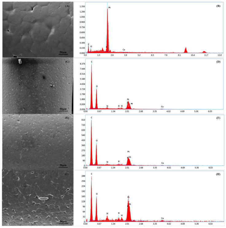
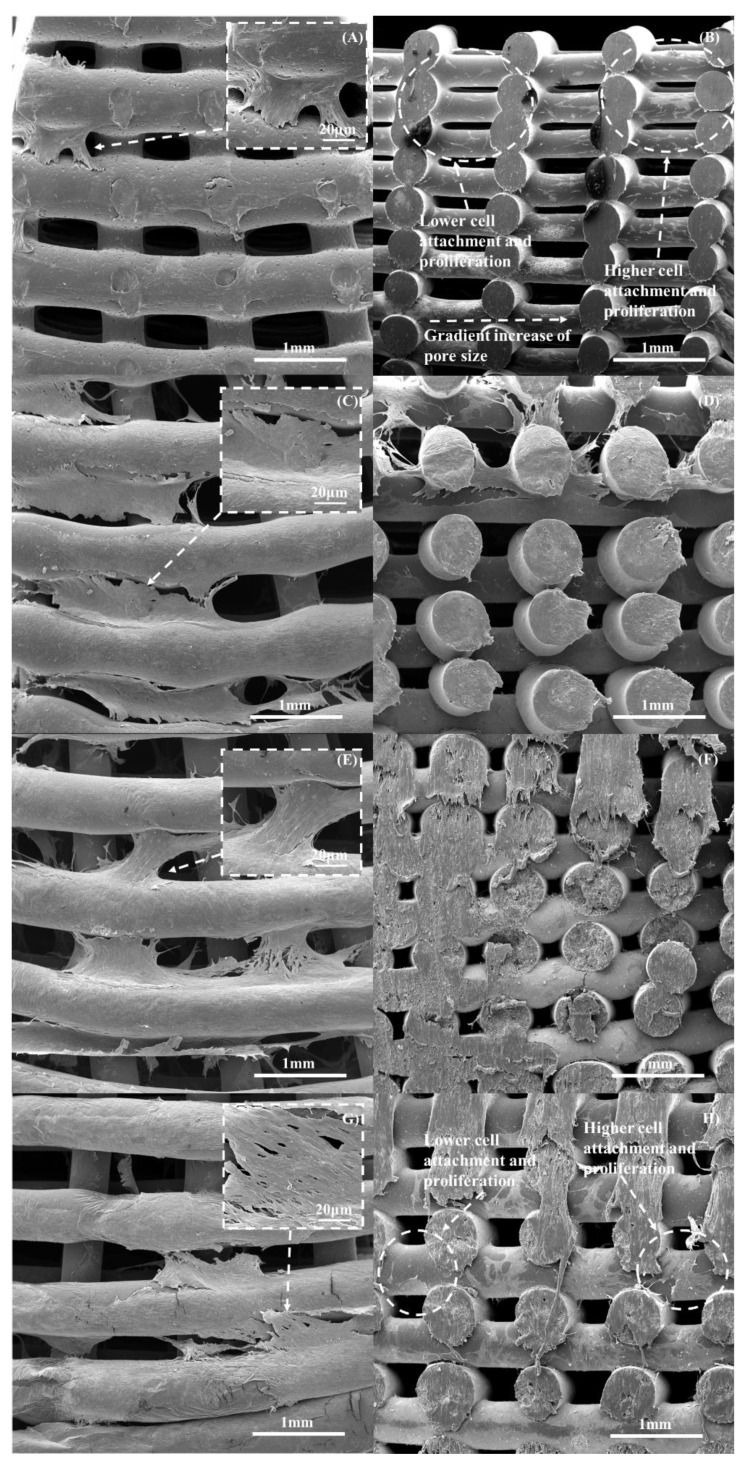
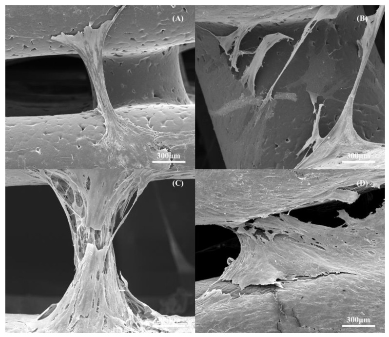
The design of scaffolds with optimal biomechanical properties for load-bearing applications is an important topic of research. Most studies have addressed this problem by focusing on the material composition and not on the coupled effect between the material composition and the scaffold architecture. Polymer-bioglass scaffolds have been investigated due to the excellent bioactivity properties of bioglass, which release ions that activate osteogenesis. However, material preparation methods usually require the use of organic solvents that induce surface modifications on the bioglass particles, compromising the adhesion with the polymeric material thus compromising mechanical properties. In this paper, we used a simple melt blending approach to produce polycaprolactone/bioglass pellets to construct scaffolds with pore size gradient. The results show that the addition of bioglass particles improved the mechanical properties of the scaffolds and, due to the selected architecture, all scaffolds presented mechanical properties in the cortical bone region. Moreover, the addition of bioglass indicated a positive long-term effect on the biological performance of the scaffolds. The pore size gradient also induced a cell spreading gradient.

Keywords: 3D printing; PCL; bioglass; bone scaffolds; tissue engineering.


Conflict of interest statement

The authors declare no conflict of interest.

Figures

Figure 1
Figure 1
The 3D Discovery system. (A) The screw and (B) the material chamber and extrusion head.
Figure 2
Figure 2
The anthropometric-based geometry and scaffolds dimensions.
Figure 3
Figure 3
The different regions for the water contact angle tests: (a) the internal region; (b) the central region; and (c) the external region.
Figure 4
Figure 4
The top view of the 10 wt% bioglass bone bricks: (A) The fabricated bone brick and (B) the SEM image exhibiting the gradient pore size.
Figure 5
Figure 5
The top and cross-section SEM images of the scaffolds with different material compositions: (A,B) PCL; (C,D) bioglass 10 wt%; (E,F) bioglass 15 wt%; and (G,H) bioglass 20 wt%.
Figure 6
Figure 6
The water drop test on a PCL scaffold filament at 0 s (A) and 20 s (B).
Figure 7
Figure 7
The Thermal Gravimetric Analysis (TGA) curves of the bioglass bone bricks.
Figure 8
Figure 8
The XRD patterns of the PCL and PCL–bioglass scaffolds (80/20 wt%) in the range of 2θ = 20–24.
Figure 9
Figure 9
The FTIR spectra of the PCL and PCL–bioglass scaffolds.
Figure 10
Figure 10
The SEM and EDX spectra of the PCL bone brick (A,B); the PCL–bioglass 10 wt% scaffolds (C,D); the PCL/HA 15 wt% scaffolds (E,F); and the PCL–bioglass 20 wt% scaffolds (G,H).
Figure 11
Figure 11
(A) The compressive modulus and (B) 0.2% offset yield strength results as a function of bioglass content. * Statistical evidence (p < 0.05) analysed by one-way ANOVA, and Tukey post hoc test. The * statistical evidence (p < 0.05), **, *** and **** is the one-way analysis of variance (one-way ANOVA) and Tukey’s post hoc test with the use of GraphPad Prism software and is used to show the difference between the results. The * is a small difference, while more * are added as the differences between the results increases.
Figure 12
Figure 12
The average fluorescence intensity for the different scaffolds at different days after cell seeding. * Statistical evidence (p < 0.05) analysed by one-way ANOVA, and Tukey post hoc test. The * Statistical evidence (p < 0.05), ** and *** is the one-way analysis of variance (one-way ANOVA) and Tukey’s post hoc test with the use of GraphPad Prism software and is used to show the difference between the results. The * is a small difference, while more * are added as the differences between the results increase.
Figure 13
Figure 13
The SEM and EDX spectra of the scaffolds at day 14 after cell seeding: PCL scaffold (A,B); PCL–bioglass 10 wt% scaffold (C,D); PCL–bioglass 15 wt% scaffold (E,F); and PCL–bioglass 20 wt% scaffold (G,H).
Figure 14
Figure 14
The cell count at day 14 after cell seeding in different regions of the scaffolds. * Statistical evidence (p < 0.05) analysed by one-way ANOVA, and Tukey post hoc test. The * statistical evidence (p < 0.05), **, *** and **** is the one-way analysis of variance (one-way ANOVA) and Tukey’s post hoc test with the use of GraphPad Prism software and is used to show the difference between the results. The * is a small difference, while more * are added as the differences between the results increases.
Figure 15
Figure 15
The top and cross-section SEM images showing cell spreading on the scaffolds with different material compositions on day 14: (A,B) PCL; (C,D) 10 wt% bioglass; (E,F) 15 wt% bioglass; and (G,H) 20 wt% bioglass.
Figure 16
Figure 16
The cell bridging between the 3D printed filaments at day 14 after cell seeding: (A) PCL scaffold; (B) PCL–bioglass 10 wt%; (C) PCL–bioglass 15 wt%; and (D) PCL–bioglass 20 wt%.

References

    1. Eivazzadeh-Keihan R., Bahojb N.E., Khanmohammadi C.K., Jafari A., Radinekiyan F., Hashemi S., Ahmadpour F., Behboudi A., Mosafer J., Mokhtarzadeh A., et al. Metal-based nanoparticles for bone tissue engineering. J. Tissue Eng. Regener. Med. 2020;14:1687–1714. doi: 10.1002/term.3131. - DOI - PubMed
    1. Wang W., Zhang B., Li M., Li J., Zhang C., Han Y., Wang L., Wang K., Zhou C., Liu L., et al. 3D printing of PLA/n-HA composite scaffolds with customized mechanical properties and biological functions for bone tissue engineering. Compos. Part B Eng. 2021;224:109192. doi: 10.1016/j.compositesb.2021.109192. - DOI
    1. Koons L.G., Diba M., Mikos G.A. Materials design for bone-tissue engineering. Nat. Rev. Mater. 2020;5:584–603. doi: 10.1038/s41578-020-0204-2. - DOI
    1. Collins N.M., Ren G., Young K., Pina S., Reis L.R., Oliveira M.J. Scaffold Fabrication Technologies and Structure/Function Properties in Bone Tissue Engineering. Adv. Funct. Mater. 2021;31:2010609. doi: 10.1002/adfm.202010609. - DOI
    1. Ulson O., Zamboni C., Durigan R.J., Hungria S.O.J., Neto J.S.H., Christian R.W., Mercadante T.M., Santili C. Treatment of femur pseudoarthrosis using wave plate: Evaluation of consolidation and its relationship with graft type. Injury. 2021;52:S18–S22. doi: 10.1016/j.injury.2021.01.045. - DOI - PubMed

LinkOut - more resources