. 2022 Jan 23;14(3):454.
doi: 10.3390/polym14030454.

Metronidazole Topically Immobilized Electrospun Nanofibrous Scaffold: Novel Secondary Intention Wound Healing Accelerator

Affiliations

Metronidazole Topically Immobilized Electrospun Nanofibrous Scaffold: Novel Secondary Intention Wound Healing Accelerator

Ahmed A El-Shanshory et al. Polymers (Basel). .

Abstract

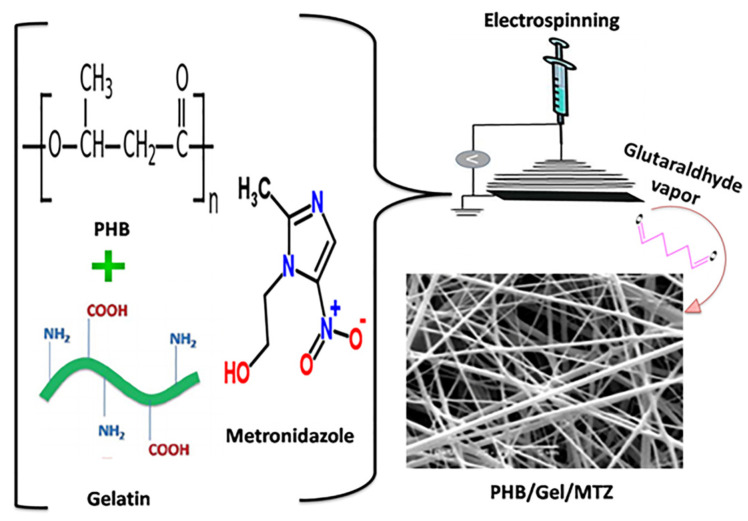
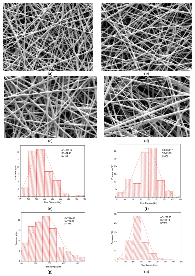
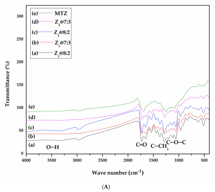
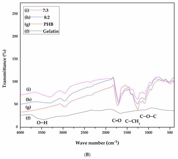
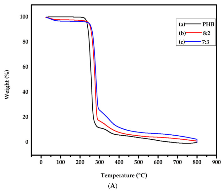
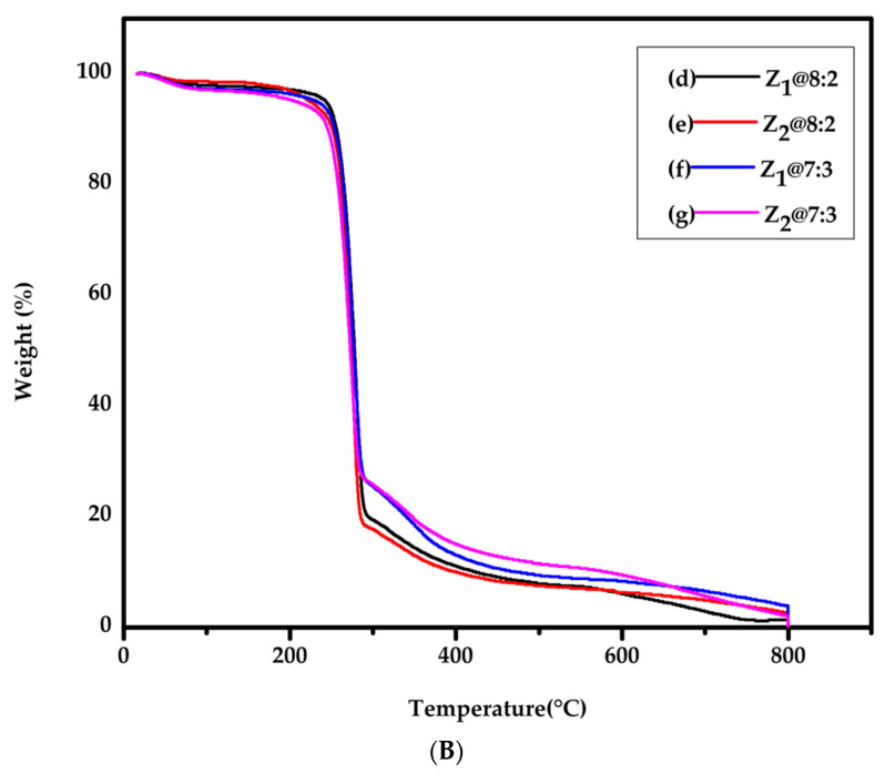
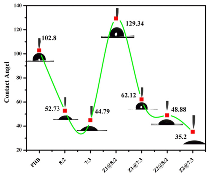
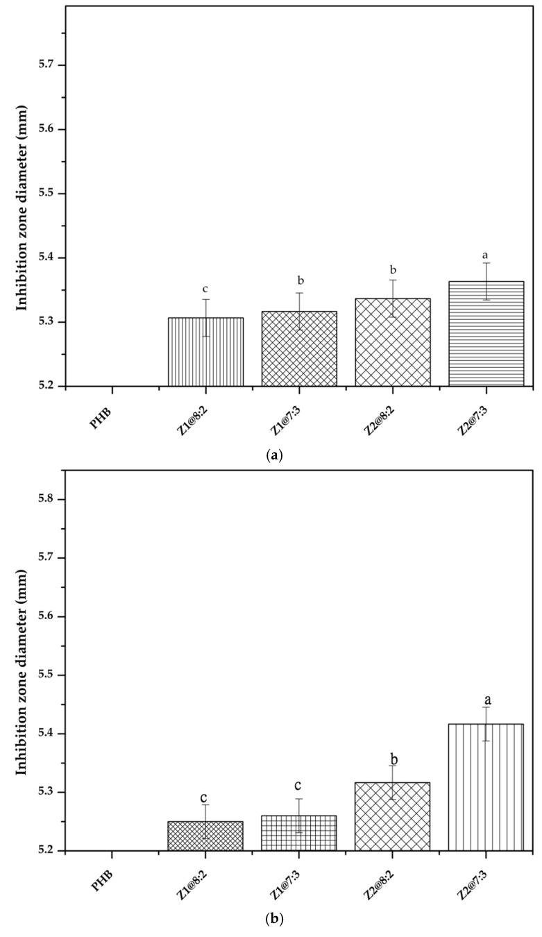
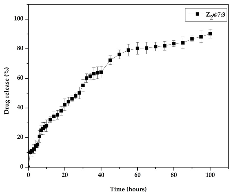
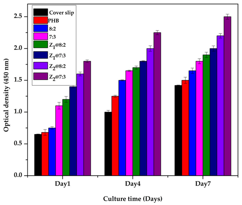
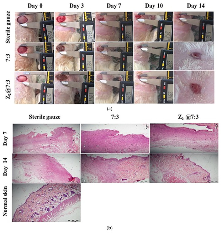
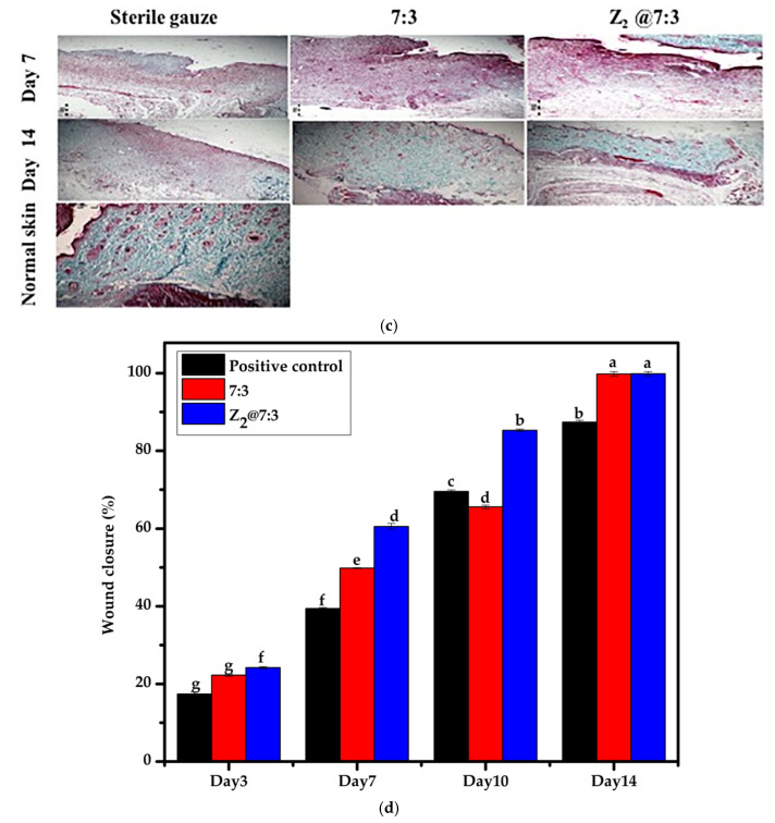
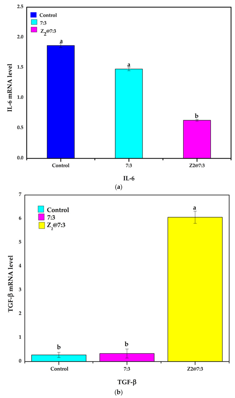
The process of secondary intention wound healing includes long repair and healing time. Electrospun nanofibrous scaffolds have shown potential for wound dressing. Biopolymers have gained much attention due to their remarkable characteristics such as biodegradability, biocompatibility, non-immunogenicity and nontoxicity. This study anticipated to develop a new composite metronidazole (MTZ) immobilized nanofibrous scaffold based on poly (3-hydroxy butyrate) (PHB) and Gelatin (Gel) to be utilized as a novel secondary intention wound healing accelerator. Herein, PHB and Gel were mixed together at different weight ratios to prepare polymer solutions with final concentration of (7%), loaded with two different concentrations 5% (Z1) and 10% (Z2) of MTZ. Nanofibrous scaffolds were obtained by manipulating electrospinning technique. The properties of MTZ immobilized PHB/Gel nanofibrous scaffold were evaluated (SEM, FTIR, TGA, water uptake, contact angle, porosity, mechanical properties and antibacterial activity). Additionally, in vitro cytocompatibility of the obtained nanofibrous scaffolds were assessed by using the cell counting kit-8 (CCK-8 assay). Moreover, in vivo wound healing experiments revealed that the prepared nanofibrous scaffold highly augmented the transforming growth factor (TGF-β) signaling pathway, moderately suppressed the pro-inflammatory cytokine (IL-6). These results indicate that MTZ immobilized PHB/Gel nanofibrous scaffold significantly boost accelerating secondary intention wound healing.

Keywords: antibacterial activity; biocompatibility; electrospinning; metronidazole; nanofibrous scaffold; secondary intention wound healing.


Conflict of interest statement

The authors declare no competing interest.

Figures

Scheme 1
Scheme 1
Schematic diagram of synthesis process and chemical structure of the molecules and polymers involved in the synthesis of nanofibrous scaffold Z2@7:3.
Figure 1
Figure 1
Nanofibrous scaffolds morphology and their relative histogram exhibiting average fiber diameter. SEM images of PHB/Gel nanofibrous scaffolds with different ratios (8:2 and 7:3) loaded with two different concentrations of MTZ 5% (Z1) (a,c) and MTZ 10% (Z2) (b,d), respectively, and histograms exhibiting average fiber diameter with SD± (eh).
Figure 2
Figure 2
FT-IR spectra of (A) MTZ, PHB/Gel with different ratios loaded with MTZ at two different concentrations Z1 and Z2.; (B) gelatin, PHB and PHB/Gel with different ratios (8:2, 7:3).
Figure 2
Figure 2
FT-IR spectra of (A) MTZ, PHB/Gel with different ratios loaded with MTZ at two different concentrations Z1 and Z2.; (B) gelatin, PHB and PHB/Gel with different ratios (8:2, 7:3).
Figure 3
Figure 3
TGA curves of (A) PHB and PHB/Gel with different ratios (8:2 and 7:3); (B) PHB/gel/MTZ (8:2 and 7:3) with two different concentrations Z1 and Z2.
Figure 3
Figure 3
TGA curves of (A) PHB and PHB/Gel with different ratios (8:2 and 7:3); (B) PHB/gel/MTZ (8:2 and 7:3) with two different concentrations Z1 and Z2.
Figure 4
Figure 4
Contact angle values of nanofibrous scaffolds PHB, 8:2, 7:3, Z1@8:2, Z1@7:3, Z2@8:2 and Z2@7:3.
Figure 5
Figure 5
Antimicrobial activity of nanofibrous scaffolds against (a) E. coli. (b) S. aureus. Different letters mean statistically significant differences for a p-value < 0.05.
Figure 6
Figure 6
In vitro release profile of MTZ from Z2@7:3 nanofibrous scaffold in PBS (pH = 7.4).
Figure 7
Figure 7
Cell biocompatibility on different nanofibrous scaffolds. L-929 cells proliferation on PHB/Gel/MTZ nanofibrous scaffolds.
Figure 8
Figure 8
(a) Photographic images of in vivo full thickness excision wounds after treatment with sterile gauze, blank and Z2@7:3 for 14 days. (b) Histological analysis of treated wounds on days 7 and 14 using H&E stain. (c) Histological analysis of treated wounds using Masson’s trichrome Stain (MTS) on day 7 and 14 (original magnification = 100). (d) Percentage of wound closure in three different groups at different time, different letters mean statistically significant differences for a p-value < 0.05.
Figure 8
Figure 8
(a) Photographic images of in vivo full thickness excision wounds after treatment with sterile gauze, blank and Z2@7:3 for 14 days. (b) Histological analysis of treated wounds on days 7 and 14 using H&E stain. (c) Histological analysis of treated wounds using Masson’s trichrome Stain (MTS) on day 7 and 14 (original magnification = 100). (d) Percentage of wound closure in three different groups at different time, different letters mean statistically significant differences for a p-value < 0.05.
Figure 9
Figure 9
Expression levels of genes related to wound healing on day 14. (a) IL-6; (b) TGF-β. Different letters mean statistically significant differences for a p-value < 0.05.

References

    1. Boer M., Duchnik E., Maleszka R., Marchlewicz M. Structural and biophysical characteristics of human skin in maintaining proper epidermal barrier function. Adv. Dermatol. Allergol. 2016;1:1–5. doi: 10.5114/pdia.2015.48037. - DOI - PMC - PubMed
    1. Khan M.A., Hussain Z., Ali S., Qamar Z., Imran M., Hafeez F.Y. Fabrication of Electrospun Probiotic Functionalized Nanocomposite Scaffolds for Infection Control and Dermal Burn Healing in a Mice Model. ACS Biomater. Sci. Eng. 2019;5:6109–6116. doi: 10.1021/acsbiomaterials.9b01002. - DOI - PubMed
    1. Xue M., Jackson C.J. Extracellular Matrix Reorganization During Wound Healing and Its Impact on Abnormal Scarring. Adv. Wound Care. 2015;4:119–136. doi: 10.1089/wound.2013.0485. - DOI - PMC - PubMed
    1. Ma L. Collagen/chitosan porous scaffolds with improved biostability for skin tissue engineering. Biomaterials. 2003;24:4833–4841. doi: 10.1016/S0142-9612(03)00374-0. - DOI - PubMed
    1. Sarhan W.A., Azzazy H.M.E., El-Sherbiny I.M. Honey/Chitosan Nanofiber Wound Dressing Enriched with Allium sativum and Cleome droserifolia: Enhanced Antimicrobial and Wound Healing Activity. ACS Appl. Mater. Interfaces. 2016;8:6379–6390. doi: 10.1021/acsami.6b00739. - DOI - PubMed

LinkOut - more resources