Review
. 2022 Jan 30;14(3):566.
doi: 10.3390/polym14030566.

Unraveling of Advances in 3D-Printed Polymer-Based Bone Scaffolds

Affiliations
Review

Unraveling of Advances in 3D-Printed Polymer-Based Bone Scaffolds

Yuanhang Xu et al. Polymers (Basel). .

Abstract

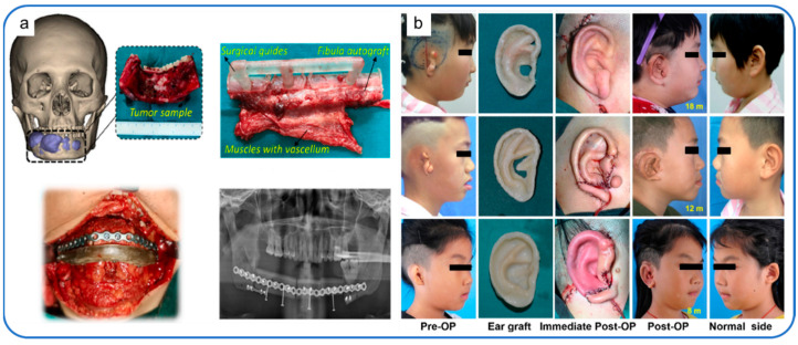
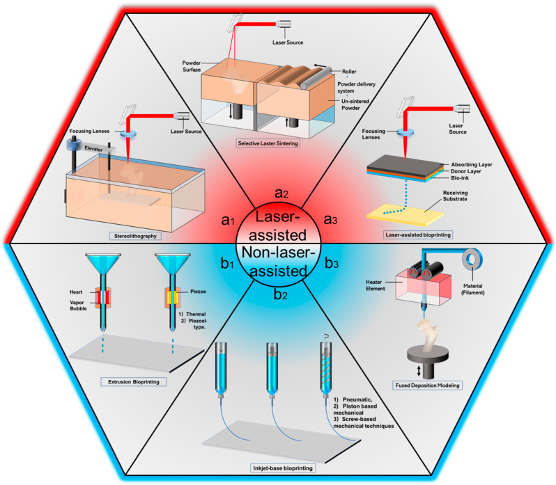
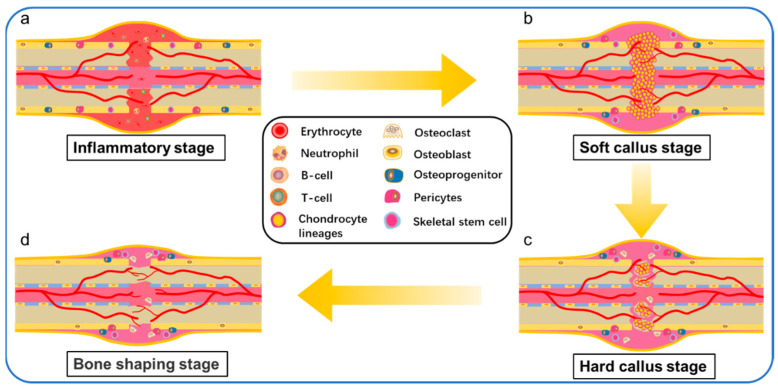
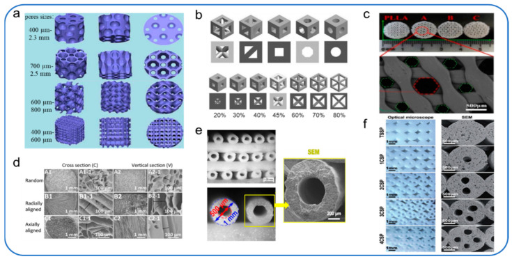
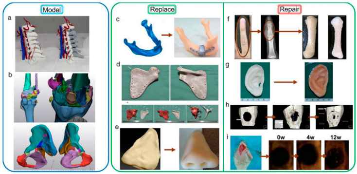
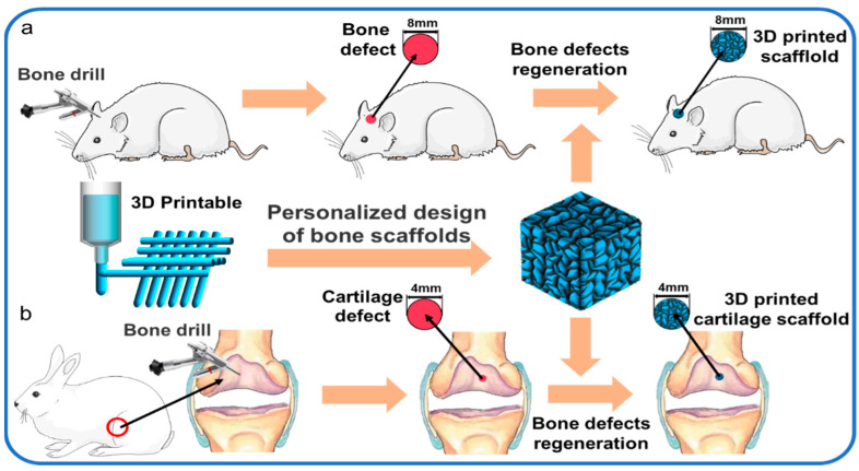
The repair of large-area irregular bone defects is one of the complex problems in orthopedic clinical treatment. The bone repair scaffolds currently studied include electrospun membrane, hydrogel, bone cement, 3D printed bone tissue scaffolds, etc., among which 3D printed polymer-based scaffolds Bone scaffolds are the most promising for clinical applications. This is because 3D printing is modeled based on the im-aging results of actual bone defects so that the printed scaffolds can perfectly fit the bone defect, and the printed components can be adjusted to promote Osteogenesis. This review introduces a variety of 3D printing technologies and bone healing processes, reviews previous studies on the characteristics of commonly used natural or synthetic polymers, and clinical applications of 3D printed bone tissue scaffolds, analyzes and elaborates the characteristics of ideal bone tissue scaffolds, from t he progress of 3D printing bone tissue scaffolds were summarized in many aspects. The challenges and potential prospects in this direction were discussed.

Keywords: 3D printing; bone healing; bone tissue engineering scaffolds; polymer.


Conflict of interest statement

The authors declare no conflict of interest.

Figures

Figure 5
Figure 5
(a) The clinical implantation and post-operative effect of 3D-printed PEEK implant; reproduced with permission from Kang, J [86], published by J Mech Behav Biomed Mater 2021, 116, 104335; (b) Composite tissue scaffold loaded with cartilage cells implanted in the human body; reproduced with permission from Zhou, G. [178], published by EBioMedicine 2018, 28, 287–302.
Figure 1
Figure 1
3D printing process diagram: (a1) SLS; (a2) SLA; (a3) LAD; (b1) Extrusion Bioprinting; (b2) Inkjet Bioprinting; (b3) FDM.
Figure 2
Figure 2
Repair processes of bone defeat. (a) Inflammatory stage; (b) Soft callus stage; (c) Hard callus stage; (d) Bone shaping stage.
Figure 3
Figure 3
Pore structure diversity of 3D printed bone scaffolds. (a) Based on the observation and analysis of lotus root structure and bone trabecular structure, four kinds of porous scaffolds were proposed; reproduced with permission from Zhipeng, H. et al. [66], published by Composites Science and Technology 2020. (b) Five pore geometries were selected (triangular prism with a rounded and a flat profile, cube, octagonal prism, sphere) and seven porosities (up to 80%), on the basis of which 70 models were created for finite element analyses;reproduced with permission from Piotr, P. et al. [68], published by Materials 2020; (c) The porous g-HNTs/g-MgOs/PLLA composite scaffolds with large and small pores and honeycomb structure; reproduced with permission from Kun, L. et al. [69], published by Composites Part B: Engineering 2020; (d) SF/COL composite scaffolds with random pores, radially aligned pores or axially aligned pores; reproduced with permission from Xue, F. et al. [70], published by Journal of Materials Chemistry B 2020; (e,f) Hollow channels structure; reproduced with permission from Wenjie, Z. et al. [71], published by Biomaterials 2017; reproduced with permission from Chun, F. et al. [72], published by Advanced Science 2017.
Figure 4
Figure 4
3D-printed for Medical Applications. (a) Preoperative full-color analysis model of a patient with C6 vertebral bone tumor; (b) Preoperative modeling of comminuted fractures in the knee and pelvis; (c) Gradient lattice mandibular prosthesis prepared by metal coated polymer; reproduced with permission from Xiao, R. [161], published by Composites Part B: Engineering 2020, 193, 108057; (d) Scapular prosthesis made with PEEK; reproduced with permission from Liu, D. [162], published by J Bone Oncol 2018, 12, 78–82; (e) Middle phalangeal bone implant prepared by sPLA/N-HAP composite; reproduced with permission from Nuseir, A. [78], published by J Prosthodont 2019, 28, 10–14; (f) Micro-ct showing BMP-2 loaded SFF stent promoting bone healing;reproduced with permission from Gupta, A. [163], published by ACS omega 2017, 2, 4039–4052; (g) human ear-shaped cartilage reconstruction based on tissue engineering scaffolds; reproduced with permission from Zhou, G. [164], published by EBioMedicine 2018, 28, 287–302; (h) Micro-CT images of bone regeneration in the defects of BMP-2-loaded SFF scaffold; reproduced with permission from Lee, J.W. [79], published by Biomaterials 2011, 32, 744–752; (i) Micro-CT images of bone regeneration in the defects of Biodegradable 3D scaffolds; reproduced with permission from Li, X. [165], published by Macromol Biosci 2018, 18, e1800068.
Figure 6
Figure 6
(a) regeneration of skull defect in rats; (b) articular cartilage regeneration in rabbits.

Similar articles

Cited by

References

    1. Tuan Rahim T.N.A., Abdullah A., Md Akil H., Mohamad D., Rajion Z. The improvement of mechanical and thermal properties of polyamide 12 3D printed parts by fused deposition modelling. Express Polym. Lett. 2017;11:963–982. doi: 10.3144/expresspolymlett.2017.92. - DOI
    1. Bao X., Zhu L., Huang X., Tang D., He D., Shi J., Xu G. 3D biomimetic artificial bone scaffolds with dual-cytokines spatiotemporal delivery for large weight-bearing bone defect repair. Sci. Rep. 2017;7:7814. doi: 10.1038/s41598-017-08412-0. - DOI - PMC - PubMed
    1. Biggemann J., Pezoldt M., Stumpf M., Greil P., Fey T. Modular ceramic scaffolds for individual implants. Acta Biomater. 2018;80:390–400. doi: 10.1016/j.actbio.2018.09.008. - DOI - PubMed
    1. Pape H.C., Evans A., Kobbe P. Autologous Bone Graft: Properties and Techniques. J. Orthop. Trauma. 2010;24:36–40. doi: 10.1097/BOT.0b013e3181cec4a1. - DOI - PubMed
    1. Larsen M., Pelzer M., Friedrich P.F., Wood C.M., Bishop A.T. Living bone allotransplants survive by surgical angiogenesis alone: Development of a novel method of composite tissue allotransplantation. J. Bone Jt. Surg. Am. 2011;93:261–273. doi: 10.2106/JBJS.G.01152. - DOI - PMC - PubMed

LinkOut - more resources