. 2022 Jan 18;15(3):711.
doi: 10.3390/ma15030711.

Clinical Evaluation of Magnesium Alloy Osteosynthesis in the Mandibular Head

Affiliations

Clinical Evaluation of Magnesium Alloy Osteosynthesis in the Mandibular Head

Marcin Kozakiewicz et al. Materials (Basel). .

Abstract

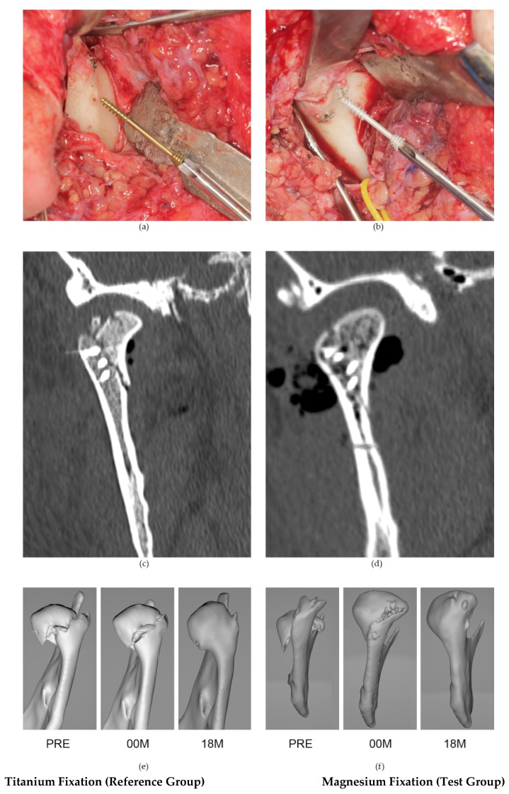
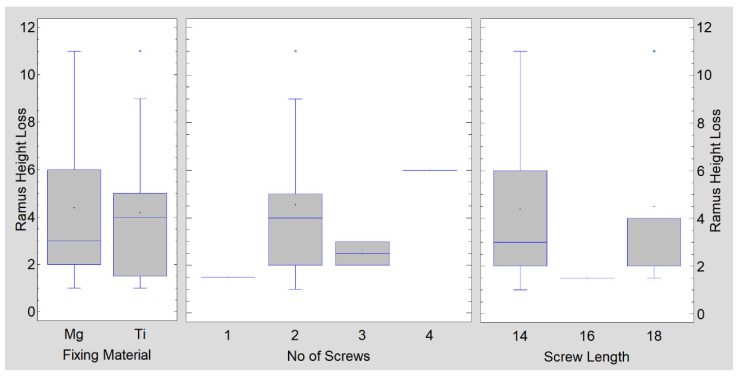
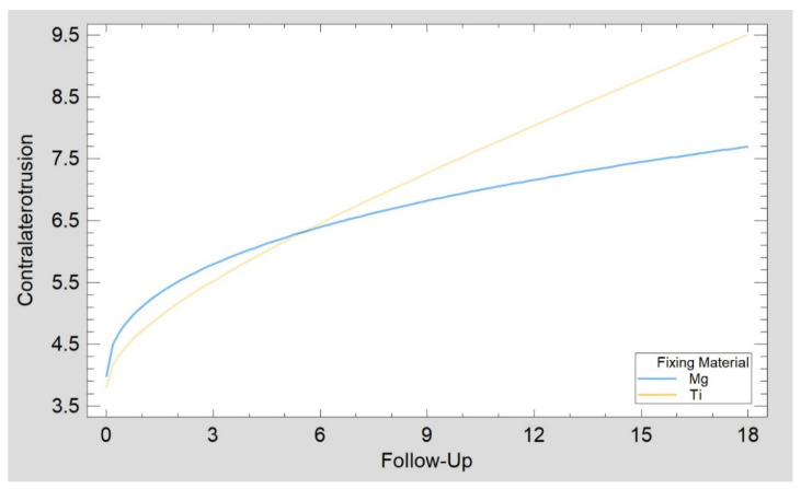
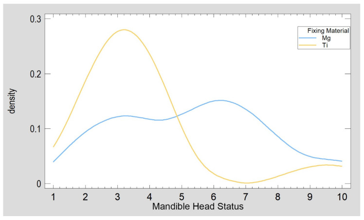
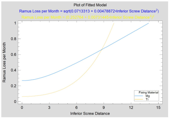
Titanium alloys are used in skeletal surgery. However, once bone union is complete, such fixation material becomes unnecessary or even harmful. Resorbable magnesium materials have been available for several years (WE43 alloy). The aim of this study was to clinically compare magnesium versus titanium open reduction and rigid fixations in mandible condylar heads. Ten patients were treated for fractures of the mandibular head with magnesium headless compression screws (2.3 mm in diameter), and 11 patients were included as a reference group with titanium screws (1.8 mm in diameter) with similar construction. The fixation characteristics (delay, time, and number of screws), distant anatomical results (mandibular ramus height loss, monthly loss rate, and relative loss of reconstructed ramus height), basic functional data (mandibular movements, facial nerve function, and cutaneous perception) and the influence of the effects of the injury (fracture type, fragmentation, occlusion, additional fractures, and associated diseases) on the outcome were evaluated. The long-term results of treatment were evaluated after 18 months. Treatment results similar to those of traditional titanium fixation were found with magnesium screws. Conclusions: Resorbable metal screws can be a favored option for osteosynthesis because surgical reentry can be avoided. These materials provide proper and stable treatment results.

Keywords: condylar head fracture; fixing material; fracture treatment; magnesium; mandible condyle; mandible fracture; mandible head; open rigid internal fixation; osteosynthesis; surgical treatment.


Conflict of interest statement

The authors declare no conflict of interest.

Figures

Figure 1
Figure 1
Examples of the clinical use of the 18 mm-long, headless compression titanium screw system 1.8 (a) and the 14 mm-long, headless compression magnesium screw system 2.3 (b) in the fixation of mandibular head fractures with a preauricular approach. Below, coronal computer tomography scans taken immediately after fixation: (c) osteosynthesis performed with three titanium screws and (d) osteosynthesis performed with four magnesium screws. Two groups for comparison: follow-up of titanium fixation on the left-hand side (reference group) and magnesium fixation on the right-hand side: (e,f): PRE-preoperative computer tomography (CT), 00 M—immediate postoperative CT, 18 M—18-month postoperative follow-up.
Figure 2
Figure 2
Method of determining mandibular ramus height (h). *—Tangent line to the posterior border of the mandibular ramus. h is determined by the length between the highest and lowest points of the ramus parallel to the tangent line.
Figure 3
Figure 3
Method for measuring the distance from the screw edge to the fracture line. Measurements were performed using CT volumetric data immediately after fixation of the bone fragments. Example of an imaging study of a patient with osteosynthesis with three magnesium screws of a type C mandibular head fracture on the right side. An asterisk (*) indicates the line tangent to the posterior border of the mandibular ramus in lateral view. Measurements of the superior (S), inferior (I), and anterior (A) screws were taken parallel to the tangent line *.
Figure 4
Figure 4
Confirmation of bone union at the fracture fixation site of the mandibular head. Regions of interest (ROIs) are described in the postfracture site in computer tomography (CT) scans taken 18 months after osteosynthesis (red square) and in cancellous control bone (green square). On the right, these areas are presented in an enlarged form (CT). Further to the right, maps of the distribution of the bone texture feature in these ROIs are shown. Texture analysis (TA) includes the sum of the squares of the optical densities acquired by CT in increments of two pixels. A finer pattern of structure can be seen in the control site than in the postfracture site, where there is normal bone interspersed with more opaque bands (p < 0.05).
Figure 5
Figure 5
As far as the mandible ramus loss is concerned, the fixing material (p = 0.879), number of used screws (p = 0.637), and length of used screws (p = 0.684) have no statistical significance.
Figure 6
Figure 6
Recovery of contralateral to fracture side range of movement (mm) as a function of time (months) was the same in patients treated with magnesium screws as in the reference group (patients treated with titanium screws). The Kolmogorov–Smirnov statistic of the regression plots (p = 0.699) confirmed similar recovery in the test group (Mg) as in the reference group (Ti).
Figure 7
Figure 7
Characteristics of patients included in this study concerning pathomorphological and pathophysiological disturbances after mandibular head injury. The groups were similar (mandible head status, p = 0.050), although there were many comminuted fractures in the test (Mg) group.
Figure 8
Figure 8
The location of the formed gas is marked in the darkest gray color. Hydrogen volume significantly decreased (p < 0.05) during the 3-month observation, i.e., between the third (a) and sixth postoperative months (b). The uniformly gray areas are an image of compact bone, while the light gray areas are an image of trabecular bone. The blue arrow marks the areas of gas inside the bone. These areas are dark gray.
Figure 9
Figure 9
Dependence of mandibular ramus height loss on the position of the inferior screw used for mandibular head fracture treatment. Both variables are given in millimeters. An increasingly lower position of the inferior screw (see “I” distance measurement in Figure 3) from the fracture line was related to an increasing rate of mandibular ramus height loss in the test group (p < 0.05), but no such relationship existed in the reference group (p = 0.196).

Similar articles

Cited by

References

    1. Dowgierd K., Borowiec M., Kozakiewicz M. Bone changes on lateral cephalograms and CBCT during treatment of maxillary narrowing using palatal osteodistraction with bone-anchored appliances. J. Cranio Maxillofac. Surg. 2018;46:2069–2081. doi: 10.1016/j.jcms.2018.08.006. - DOI - PubMed
    1. Dowgierd K., Larysz D., Szymor P., Kozakiewicz M. Alterations of upper airway volume caused by Le Fort III osteodistraction in children. J. Cranio Maxillofac. Surg. 2020;48:825–831. doi: 10.1016/j.jcms.2020.06.007. - DOI - PubMed
    1. Dowgierd K., Pokrowiecki R., Borowiec M., Kozakiewicz M., Smyczek D., Krakowczyk Ł. A Protocol for the Use of a Combined Microvascular Free Flap with Custom-Made 3D-Printed Total Temporomandibular Joint (TMJ) Prosthesis for Mandible Reconstruction in Children. Appl. Sci. 2021;11:2176. doi: 10.3390/app11052176. - DOI
    1. Herbert T.J., Fisher W.E. Management of the fractured scaphoid using a new bone screw. J. Bone Jt. Surg. Br. 1984;66:114–123. doi: 10.1302/0301-620X.66B1.6693468. - DOI - PubMed
    1. Pavlychuk T., Chernogorskyi D., Chepurnyi Y., Neff A., Kopchak A. Application of CAD/CAM technology for surgical treatment of condylar head fractures: A preliminary study. J. Oral Biol. Craniofac. Res. 2020;10:608–614. doi: 10.1016/j.jobcr.2020.08.018. - DOI - PMC - PubMed

LinkOut - more resources