. 2022 Jan 21;15(3):800.
doi: 10.3390/ma15030800.

Poly-Lysine Dendritic Nanocarrier to Target Epidermal Growth Factor Receptor Overexpressed Breast Cancer for Methotrexate Delivery

Affiliations

Poly-Lysine Dendritic Nanocarrier to Target Epidermal Growth Factor Receptor Overexpressed Breast Cancer for Methotrexate Delivery

Pratibha Narayanan et al. Materials (Basel). .

Abstract

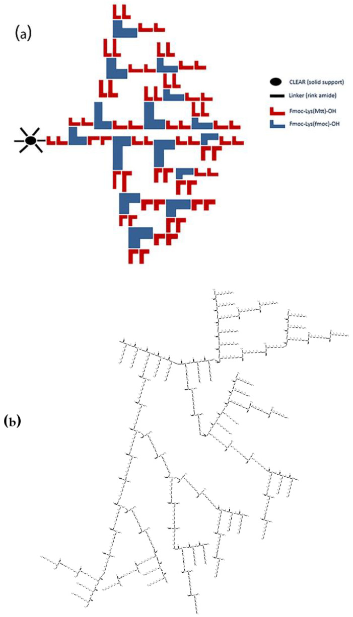
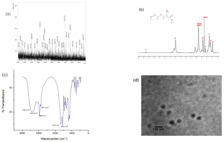
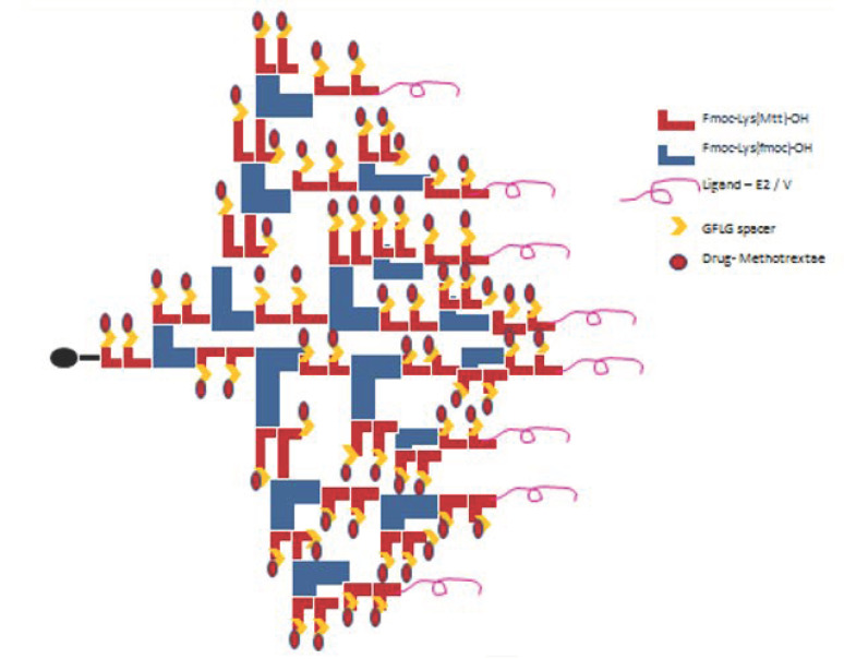
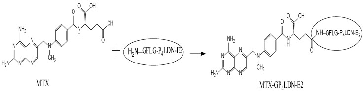
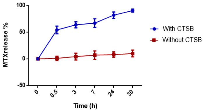
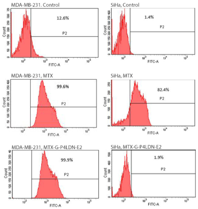
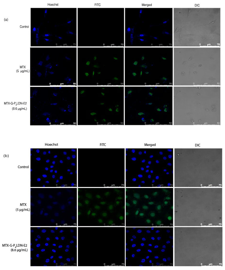
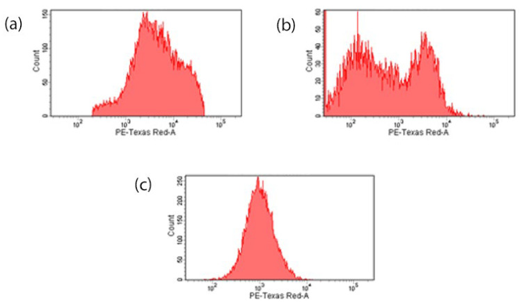
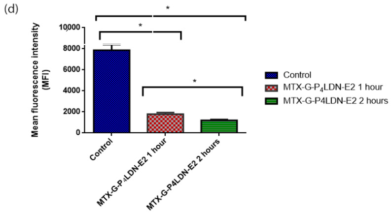
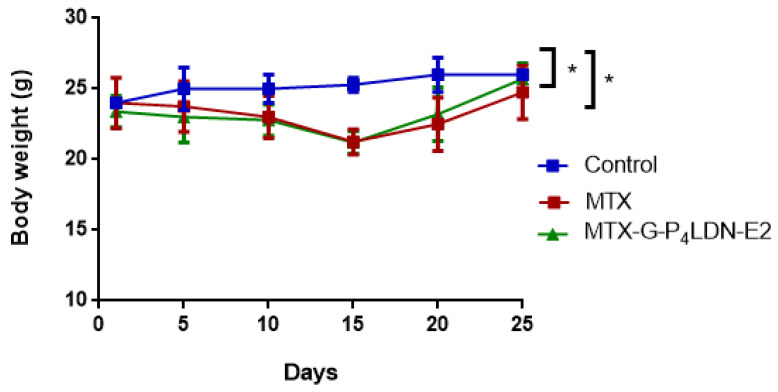
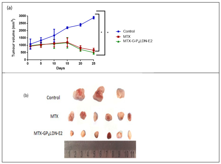
A fourth generation poly-lysine dendritic nanocarrier (P4LDN)-based targeted chemotherapy for breast cancer is attempted by incorporating an epidermal growth factor receptor (EGFR)-specific short peptide E2 (ARSHVGYTGAR) and the drug methotrexate (MTX) into a nanocarrier system. The drug is incorporated into the nanocarrier using a cathepsin B cleavable spacer: glycine-phenylalanine-leucine-glycine (GFLG). The in vitro analysis of the time-dependent drug release, binding and internalization ability, and the cytotoxic nature showed that this drug delivery system (DDS) is highly effective. The efficacy analysis using non-obese diabetic/severe combined immunodeficiency (NOD-SCID) mice also showed that compared to the control group, the DDS can effectively reduce tumor volume. The mice that received the DDS appeared to gain weight more rapidly than the free drug, which suggests that the dendrimer is more easily tolerated by mice than the free drug.

Keywords: breast cancer; epidermal growth factor receptor; in silico approach; in vivo evaluation; methotrexate; poly-lysine dendritic nanocarrier; solid-phase peptide synthesis; targeted drug delivery; tetrapeptide spacer.


Conflict of interest statement

The authors have no conflict of interest to declare.

Figures

Scheme 1
Scheme 1
(a) Synthesis protocol (attachment of lysine units is shown only in one functional site of the linker); (b) chemical structure of P4LDN after cleavage from the resin.
Figure 1
Figure 1
(a) MALDI-TOF MS profile: The [M+H]+ value calculated for C456H1030N132O63 was 9372.86 Da and found to be 9373.613 Da; (b) NMR spectra (400 MHz, DMSO-d6), ppm: 0.911 (s, 24H, NH), 1.343, 1.538, 2.253 (m, 154H, CH2), 2.762 (m, 51H, CH2–NH2), 4.204 (s, 21H, CH), 7.943, 8.232, 8.599 (t, 80H, NH2), and 13.584 (s, 2H, NH2); (c) FTIR spectra: wavenumber = 3453.5 cm−1 (m), 3308.5 cm−1 (m), 2960.6 cm−1 (m), 2871.8 cm−1 (m), 1670.7 cm−1 (s), and 1565.4 cm−1 (s); (d) TEM image of P4LDN.
Scheme 2
Scheme 2
Attachment of EGFR-targeting peptide ligand (E2), GFLG spacer (G), and the drug MTX to P4LDN.
Scheme 3
Scheme 3
Attachment of MTX to G–P4LDN–E2.
Figure 2
Figure 2
(a) MALDI-TOF MS profile: The [M+H]+ value calculated for C547H1147N163O83 was 11,336.96 Da and found to be 11,337.344 Da; (b) hydrodynamic size distribution pattern; (c) TEM image of MTX–G–P4LDN–E2 (scale bar: 100 nm).
Figure 2
Figure 2
(a) MALDI-TOF MS profile: The [M+H]+ value calculated for C547H1147N163O83 was 11,336.96 Da and found to be 11,337.344 Da; (b) hydrodynamic size distribution pattern; (c) TEM image of MTX–G–P4LDN–E2 (scale bar: 100 nm).
Figure 3
Figure 3
Time-dependent release of MTX from MTX–G–P4LDN–E2. The experiment was conducted in duplicate, and the data are represented as the arithmetic mean ± SD.
Figure 4
Figure 4
Binding abilities of MTX and MTX–G–P4LDN–E2 after 24 h of incubation with MDA-MB-231 (EGFR overexpressed) and SiHa (EGFR negative) cells. The cells without any treatment with MTX or the MTX-conjugated dendritic nanocarrier system were taken as the control.
Figure 5
Figure 5
Confocal microscopy images of (a) MDA-MB-231 (EGFR overexpressed) and (b) SiHa (EGFR negative) cells after 24 h incubation with MTX and MTX–G–P4LDN–E2.
Figure 6
Figure 6
Internalization assay: (a) control cells without a nanocarrier system; (b) 1 h incubation of MTX–G–P4LDN–E2; (c) 2 h incubation of MTX–G–P4LDN–E2 with cells; (d) mean fluorescence intensity of cells. The experiments were conducted in duplicate, and the data are represented as the arithmetic mean ± SD. The statistical significance was measured using one-way ANOVA followed by Tukey’s test (* p < 0.005).
Figure 6
Figure 6
Internalization assay: (a) control cells without a nanocarrier system; (b) 1 h incubation of MTX–G–P4LDN–E2; (c) 2 h incubation of MTX–G–P4LDN–E2 with cells; (d) mean fluorescence intensity of cells. The experiments were conducted in duplicate, and the data are represented as the arithmetic mean ± SD. The statistical significance was measured using one-way ANOVA followed by Tukey’s test (* p < 0.005).
Figure 7
Figure 7
Cytotoxicity analysis of DDS by MTT assay: (a) MDA-MB-231 cells and (b) SiHa cells. The experiment was conducted in triplicate, and the data are represented as the arithmetic mean ± SD.
Figure 8
Figure 8
Cell cycle and cell death analysis by FACS in MDA-MB-231(EGFR overexpressed) and SiHa (EGFR negative) cells after 24 h incubation with MTX and MTX–G–P4LDN–E2.
Figure 9
Figure 9
Effect of tumor treatment on body weight in female NOD-SCID mice (n = 3 for control group, n = 6 for MTX, and n = 6 for MTX–G–P4LDN–E2). The data are represented as the arithmetic mean ± SD. Statistical analysis was performed using one-way ANOVA followed by Dunnett’s test (* p < 0.05 between control and treatment groups).
Figure 10
Figure 10
(a) Anti-tumor efficacy of MTX (20 mg/kg) and MTX–G–P4LDN–E2 (35 mg/kg) in female NOD-SCID mice (n = 3 for the control group, n = 6 for MTX, and n = 6 for MTX–G–P4LDN–E2). Data are represented as the arithmetic mean ± SD, and one-way ANOVA followed by Dunnett’s test were performed for statistical analysis (* p < 0.005). (b) Comparison of tumor volumes between the control and treatment groups.

Similar articles

Cited by

  • A Recent Review on Cancer Nanomedicine.
    Giri PM, Banerjee A, Layek B. Giri PM, et al. Cancers (Basel). 2023 Apr 12;15(8):2256. doi: 10.3390/cancers15082256. Cancers (Basel). 2023. PMID: 37190185 Free PMC article. Review.
  • A DFX-based iron nanochelator for cancer therapy.
    Liu P, Wang Q, Li K, Bi B, Wen YF, Qiu MJ, Zhao J, Li BB, Zhang CH, He YL. Liu P, et al. Front Bioeng Biotechnol. 2022 Nov 28;10:1078137. doi: 10.3389/fbioe.2022.1078137. eCollection 2022. Front Bioeng Biotechnol. 2022. PMID: 36518196 Free PMC article.

References

    1. Gewirtz D.A., Bristol M.L., Yalowich J.C. Toxicity issues in cancer drug development. Curr. Opin. Investig. Drugs. 2010;11:612–614. - PubMed
    1. Boyd B.J., Kaminskas L.M., Karellas P., Krippner G., Lessene R., Porter C.J. Cationic Poly-l-lysine Dendrimers: Pharmacokinetics, Biodistribution, and Evidence for Metabolism and Bioresorption after Intravenous Administration to Rats. Mol. Pharm. 2006;3:614–627. doi: 10.1021/mp060032e. - DOI - PubMed
    1. Wang S., Chen R. pH-Responsive, Lysine-Based, Hyperbranched Polymers Mimicking Endosomolytic Cell-Penetrating Peptides for Efficient Intracellular Delivery. Chem. Mater. 2017;29:5806–5815. doi: 10.1021/acs.chemmater.7b00054. - DOI
    1. Hynes N.E., Lane H.A. ERBB receptors and cancer: The complexity of targeted inhibitors. Nat. Rev. Cancer. 2005;5:341–354. doi: 10.1038/nrc1609. - DOI - PubMed
    1. Herbst R.S., Shin D.M. Monoclonal antibodies to target epidermal growth factor receptor–positive tumors. Cancer. 2002;94:1593–1611. doi: 10.1002/cncr.10372. - DOI - PubMed