

A New Anorganic Equine Bone Substitute for Oral Surgery: Structural Characterization and Regenerative Potential
- PMID: 35160976
- PMCID: PMC8840601
- DOI: 10.3390/ma15031031
A New Anorganic Equine Bone Substitute for Oral Surgery: Structural Characterization and Regenerative Potential
Abstract
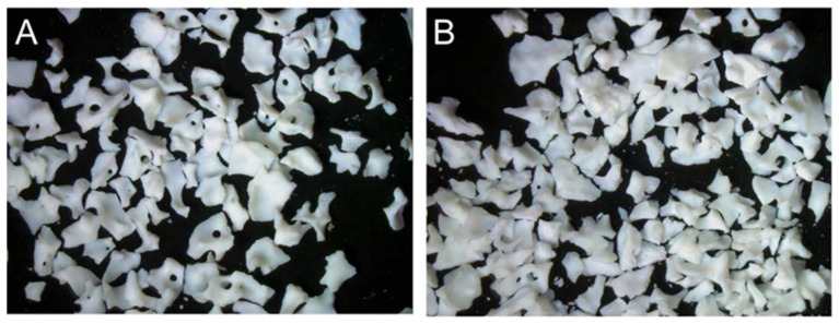
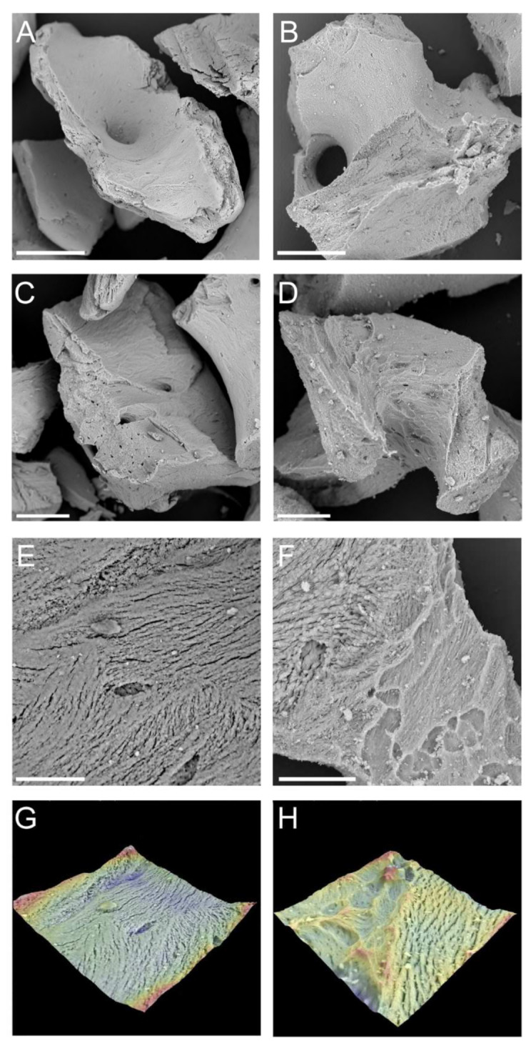
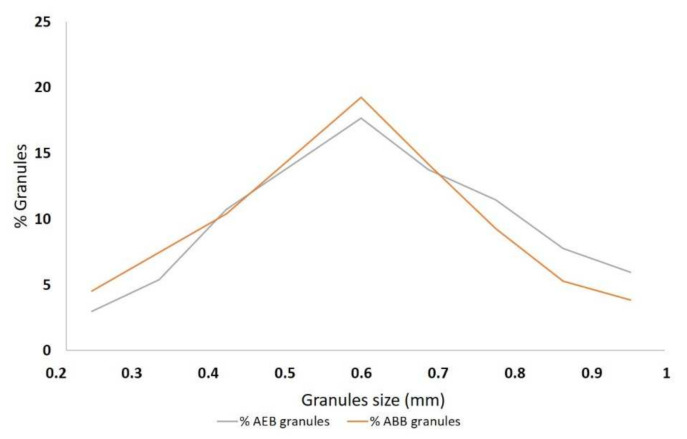
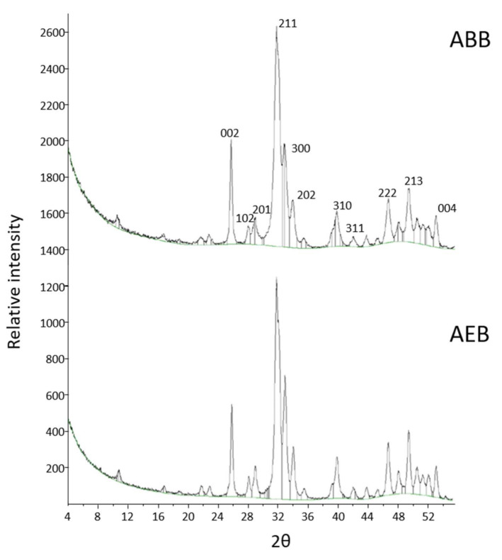
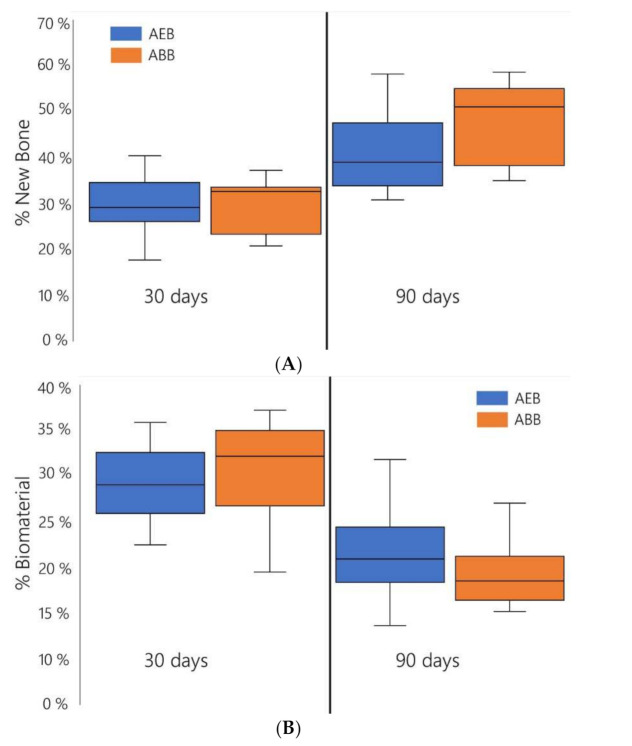
Different xenogeneic inorganic bone substitutes are currently used as bone grafting materials in oral and maxillo-facial surgery. The aim of the present study was to determine the physicochemical properties and the in vivo performance of an anorganic equine bone (AEB) substitute. AEB is manufactured by applying a process involving heating at >300 °C with the aim of removing all the antigens and the organic components. AEB was structurally characterized by scanning electron microscopy (SEM), X-ray diffraction (XRD), X-ray fluorescence (XRF), and Fourier-transformed infrared (FT-IR) spectroscopy and compared to the anorganic bovine bone (ABB). In order to provide a preliminary evaluation of the in vivo performance of AEB, 18 bone defects were prepared and grafted with AEB (nine sites), or ABB (nine sites) used as a control, in nine Yucatan Minipigs. De novo bone formation, residual bone substitute, as well as local inflammatory and tissue effects were histologically evaluated at 30 and 90 days after implantation. The structural characterization showed that the surface morphology, particle size, chemical composition, and crystalline structure of AEB were similar to cancellous human bone. The histological examination of AEB showed a comparable pattern of newly formed bone and residual biomaterial to that of ABB. Overall, the structural data and pre-clinical evidence reported in the present study suggests that AEB can be effectively used as bone grafting material in oral surgery procedures.
Keywords: anorganic bone; bone formation; equine bone substitute; xenograft.
Conflict of interest statement
Matteo Colombo, Daniele Recupero, Christian Frigerio, and Marco Morroni work for Bioteck S.p.A..
Figures
References
-
- Russmueller G., Winkler L., Lieber R., Seemann R., Pirklbauer K., Perisanidis C., Kapeller B., Spassova E., Halwax E., Poeschl W.P., et al. In Vitro effects of particulate bone substitute materials on the resorption activity of human osteoclasts. Eur. Cells Mater. 2017;34:291–306. doi: 10.22203/eCM.v034a18. - DOI - PubMed
