

SARS-CoV-2 Detection Using Optical Fiber Based Sensor Method
- PMID: 35161497
- PMCID: PMC8839674
- DOI: 10.3390/s22030751
SARS-CoV-2 Detection Using Optical Fiber Based Sensor Method
Abstract
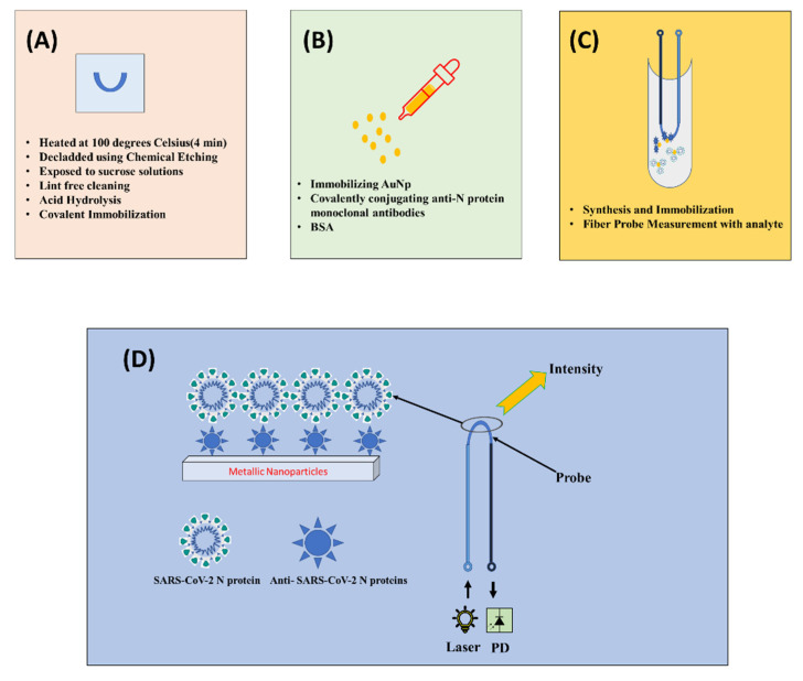
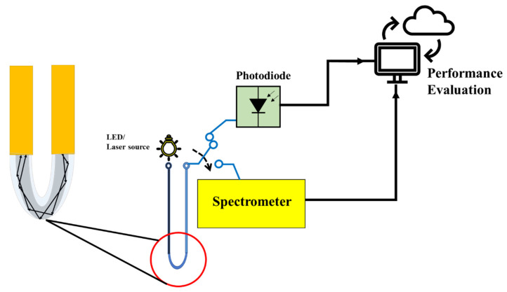
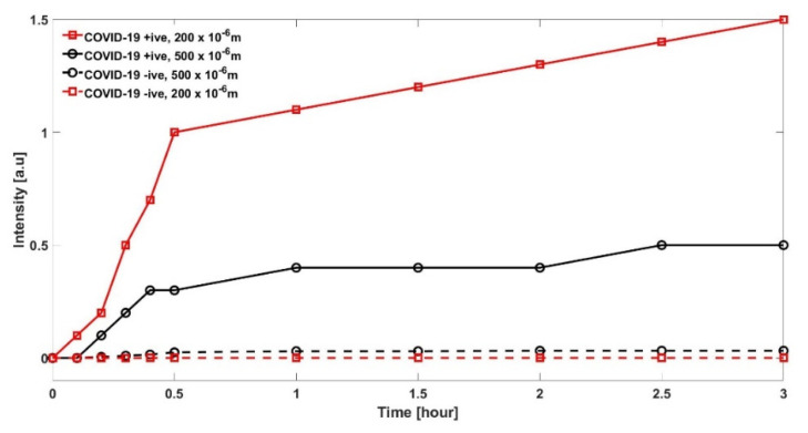
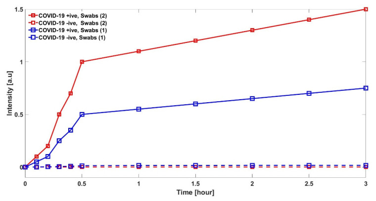
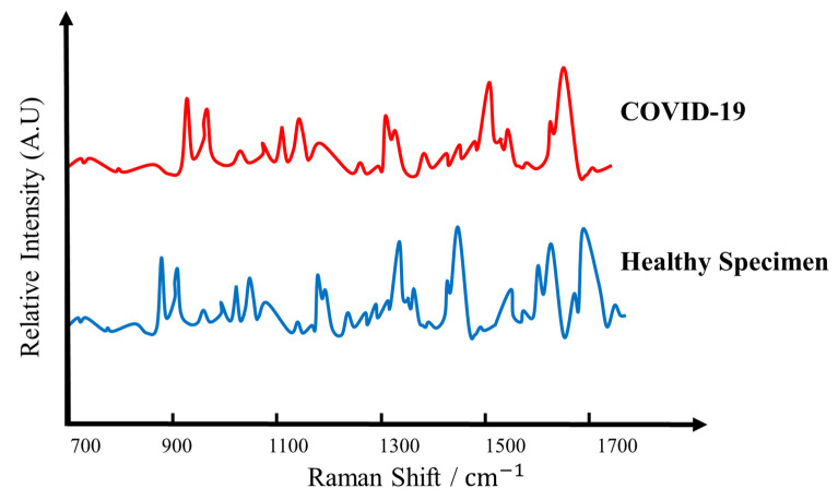
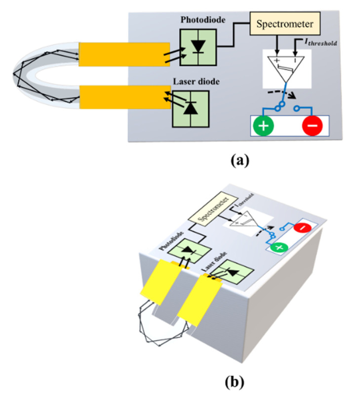
The SARS-CoV-2 Coronavirus disease, also known as the COVID-19 pandemic, has engendered the biggest challenge to human life for the last two years. With a rapid increase in the spread of the Omicron variant across the world, and to contain the spread of COVID-19 in general, it is crucial to rapidly identify this viral infection with minimal logistics. To achieve this, a novel plastic optical fiber (POF) U-shaped probe sensing method is presented for accurate detection of SARS-CoV-2, commonly known as the COVID-19 virus, which has the capability to detect new variants such as Omicron. The sample under test can be taken from oropharyngeal or nasopharyngeal via specific POF U-shaped probe with one end that is fed with a laser source while the other end is connected to a photodetector to receive the response and postprocess for decision-making. The study includes detection comparison with two types of POF with diameters of 200 and 500 µm. Results show that detection is better when a smaller-diameter POF is used. It is also seen that the proposed test bed and its envisaged prototype can detect the COVID-19 variants within 15 min of the test. The proposed approach will make the clinical diagnosis faster, cheaper and applicable to patients in remote areas where there are no hospitals or clinical laboratories due to poverty, geographic obstacles, or other factors.
Keywords: COVID-19; COVID-19 detection; PCR; SARS-CoV-2; U-shaped probe; optical fiber sensor.
Conflict of interest statement
The authors declare that they have no known competing financial interests that could have appeared to influence the work reported in this paper.
Figures
Similar articles
-
Safety and Efficacy of Imatinib for Hospitalized Adults with COVID-19: A structured summary of a study protocol for a randomised controlled trial.Trials. 2020 Oct 28;21(1):897. doi: 10.1186/s13063-020-04819-9. Trials. 2020. PMID: 33115543 Free PMC article.
-
Emergency SARS-CoV-2 Variants of Concern: Novel Multiplex Real-Time RT-PCR Assay for Rapid Detection and Surveillance.Microbiol Spectr. 2022 Feb 23;10(1):e0251321. doi: 10.1128/spectrum.02513-21. Epub 2022 Feb 23. Microbiol Spectr. 2022. PMID: 35196812 Free PMC article.
-
FnCas9-based CRISPR diagnostic for rapid and accurate detection of major SARS-CoV-2 variants on a paper strip.Elife. 2021 Jun 9;10:e67130. doi: 10.7554/eLife.67130. Elife. 2021. PMID: 34106048 Free PMC article.
-
SARS-CoV-2 variants and COVID-19 vaccines: Current challenges and future strategies.Int Rev Immunol. 2023;42(6):393-414. doi: 10.1080/08830185.2022.2079642. Epub 2022 May 28. Int Rev Immunol. 2023. PMID: 35635216 Review.
-
Revolutionizing SARS-CoV-2 omicron variant detection: Towards faster and more reliable methods.Talanta. 2024 Jan 1;266(Pt 1):124937. doi: 10.1016/j.talanta.2023.124937. Epub 2023 Jul 12. Talanta. 2024. PMID: 37481886 Review.
Cited by
-
Fibrous aggregates: Amplifying aggregation-induced emission to boost health protection.Biomaterials. 2022 Aug;287:121666. doi: 10.1016/j.biomaterials.2022.121666. Epub 2022 Jul 4. Biomaterials. 2022. PMID: 35835002 Free PMC article.
-
Rapid detection of vaccinia virus using biofunctionalized fiber-optic ball-tip biosensors.Sci Rep. 2023 Oct 14;13(1):17470. doi: 10.1038/s41598-023-44926-6. Sci Rep. 2023. PMID: 37838808 Free PMC article.
-
Rapid Optical Biosensing of SARS-CoV-2 Spike Proteins in Artificial Samples.Sensors (Basel). 2022 May 16;22(10):3768. doi: 10.3390/s22103768. Sensors (Basel). 2022. PMID: 35632177 Free PMC article.
-
Bio-Tailored Sensing at the Nanoscale: Biochemical Aspects and Applications.Sensors (Basel). 2023 Jan 13;23(2):949. doi: 10.3390/s23020949. Sensors (Basel). 2023. PMID: 36679744 Free PMC article. Review.
-
Development of an Immunocapture-Based Polymeric Optical Fiber Sensor for Bacterial Detection in Water.Polymers (Basel). 2024 Mar 21;16(6):861. doi: 10.3390/polym16060861. Polymers (Basel). 2024. PMID: 38543466 Free PMC article.
References
-
- World Health Organization . COVID-19 Weekly Epidemiological Update. World Health Organization; Geneva, Switzerland: 2021. pp. 1–3.
-
- HHS Supports Development of First High-Throughput COVID-19 Diagnostic Test. [(accessed on 21 March 2020)]; Available online: www.hhs.gov/about/news/2020/03/09/hhs-supports-development-of-firsthigh-....
MeSH terms
Supplementary concepts
LinkOut - more resources
Full Text Sources
Medical
Miscellaneous
