

E-TBNet: Light Deep Neural Network for Automatic Detection of Tuberculosis with X-ray DR Imaging
- PMID: 35161567
- PMCID: PMC8840569
- DOI: 10.3390/s22030821
E-TBNet: Light Deep Neural Network for Automatic Detection of Tuberculosis with X-ray DR Imaging
Abstract
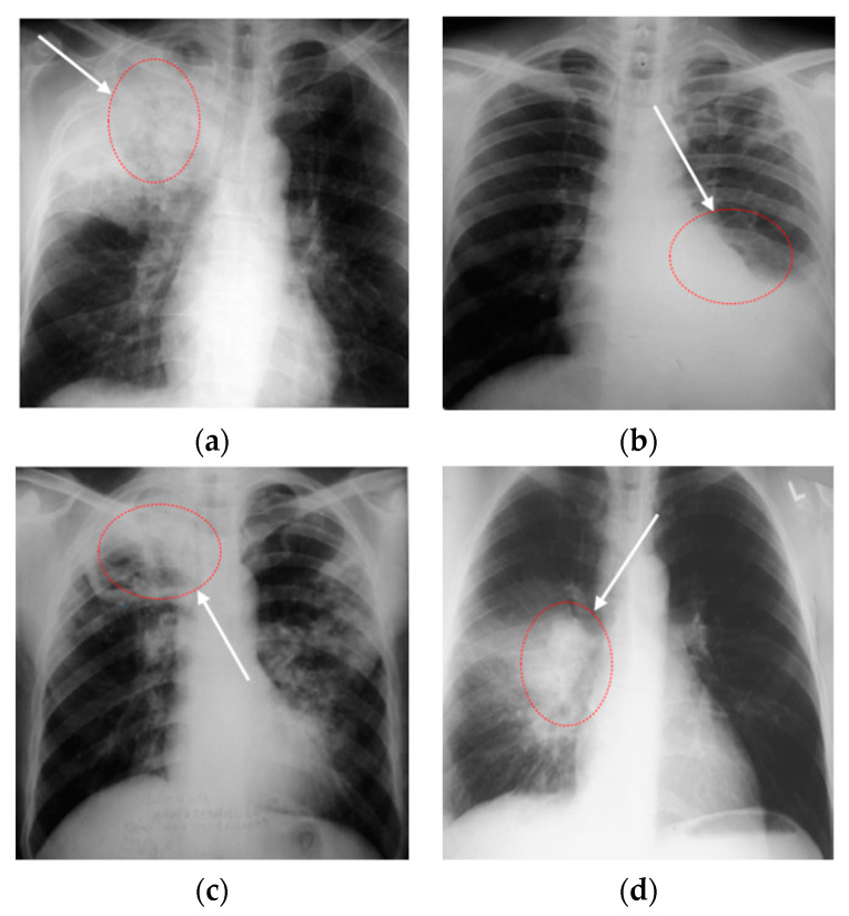
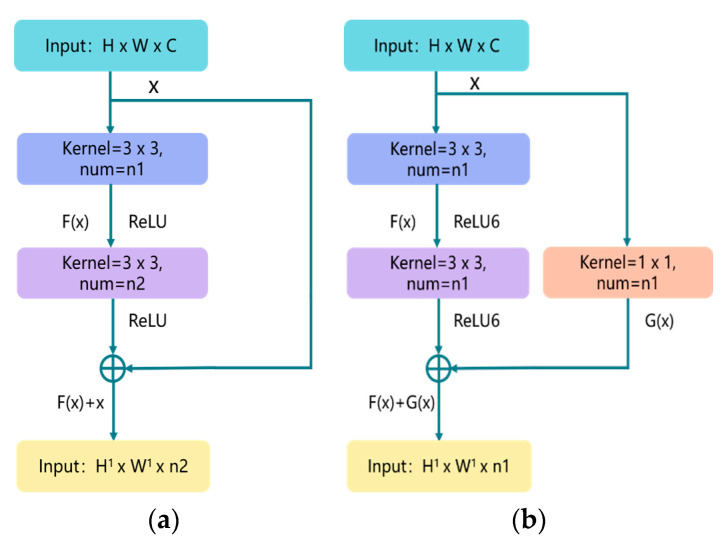
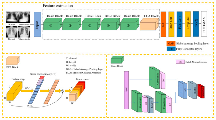
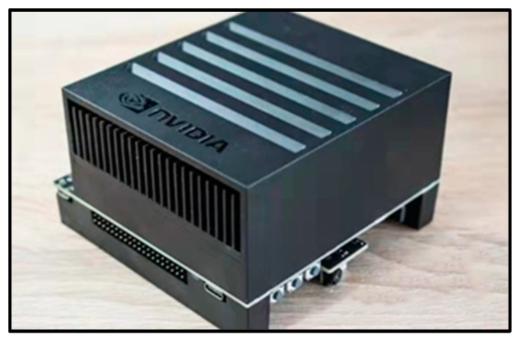
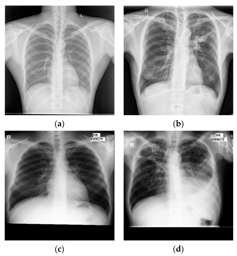
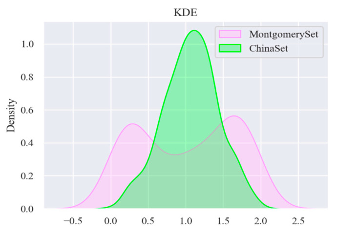
Currently, the tuberculosis (TB) detection model based on chest X-ray images has the problem of excessive reliance on hardware computing resources, high equipment performance requirements, and being harder to deploy in low-cost personal computer and embedded devices. An efficient tuberculosis detection model is proposed to achieve accurate, efficient, and stable tuberculosis screening on devices with lower hardware levels. Due to the particularity of the chest X-ray images of TB patients, there are fewer labeled data, and the deep neural network model is difficult to fully train. We first analyzed the data distribution characteristics of two public TB datasets, and found that the two-stage tuberculosis identification (first divide, then classify) is insufficient. Secondly, according to the particularity of the detection image(s), the basic residual module was optimized and improved, and this is regarded as a crucial component of this article's network. Finally, an efficient attention mechanism was introduced, which was used to fuse the channel features. The network architecture was optimally designed and adjusted according to the correct and sufficient experimental content. In order to evaluate the performance of the network, it was compared with other lightweight networks under personal computer and Jetson Xavier embedded devices. The experimental results show that the recall rate and accuracy of the E-TBNet proposed in this paper are better than those of classic lightweight networks such as SqueezeNet and ShuffleNet, and it also has a shorter reasoning time. E-TBNet will be more advantageous to deploy on equipment with low levels of hardware.
Keywords: chest X-ray images; embedded device; neural network; tuberculosis detection.
Conflict of interest statement
The authors declare no conflict of interest. The funders had no role in the design of the study; in the collection, analyses, or interpretation of data; in the writing of the manuscript; nor in the decision to publish the results. All authors read and approved the final manuscript.
Figures
References
-
- World Health Organization Global Tuberculosis Report. 2020. [(accessed on 14 October 2020)]. Available online: https://apps.who.int/iris/handle/10665/336069.
-
- Munadi K., Muchtar K., Maulina N., Pradhan B. Image Enhancement for Tuberculosis Detection Using Deep Learning. IEEE Access. 2020;8:217897–217907. doi: 10.1109/ACCESS.2020.3041867. - DOI
-
- Liu Y., Wu Y.H., Ban Y., Wang H., Cheng M.M. Rethinking Computer-Aided Tuberculosis Diagnosis; Proceedings of the Conference on Computer Vision and Pattern Recognition (CVPR 2020); Seattle, WA, USA. 13–19 June 2020; pp. 2643–2652.
-
- Rahman T., Khandakar A., Kadir M.A., Islam K.R., Islam K.F., Mazhar R., Hamid T., Islam M.T., Kashem S., Mahbub Z.B., et al. Reliable Tuberculosis Detection Using Chest X-Ray With Deep Learning, Segmentation and Visualization. IEEE Access. 2020;8:191586–191601. doi: 10.1109/ACCESS.2020.3031384. - DOI
MeSH terms
LinkOut - more resources
Full Text Sources
Medical
