

Ex Vivo Evaluation of Mechanical Anisotropic Tissues with High-Frequency Ultrasound Shear Wave Elastography
- PMID: 35161728
- PMCID: PMC8838528
- DOI: 10.3390/s22030978
Ex Vivo Evaluation of Mechanical Anisotropic Tissues with High-Frequency Ultrasound Shear Wave Elastography
Abstract
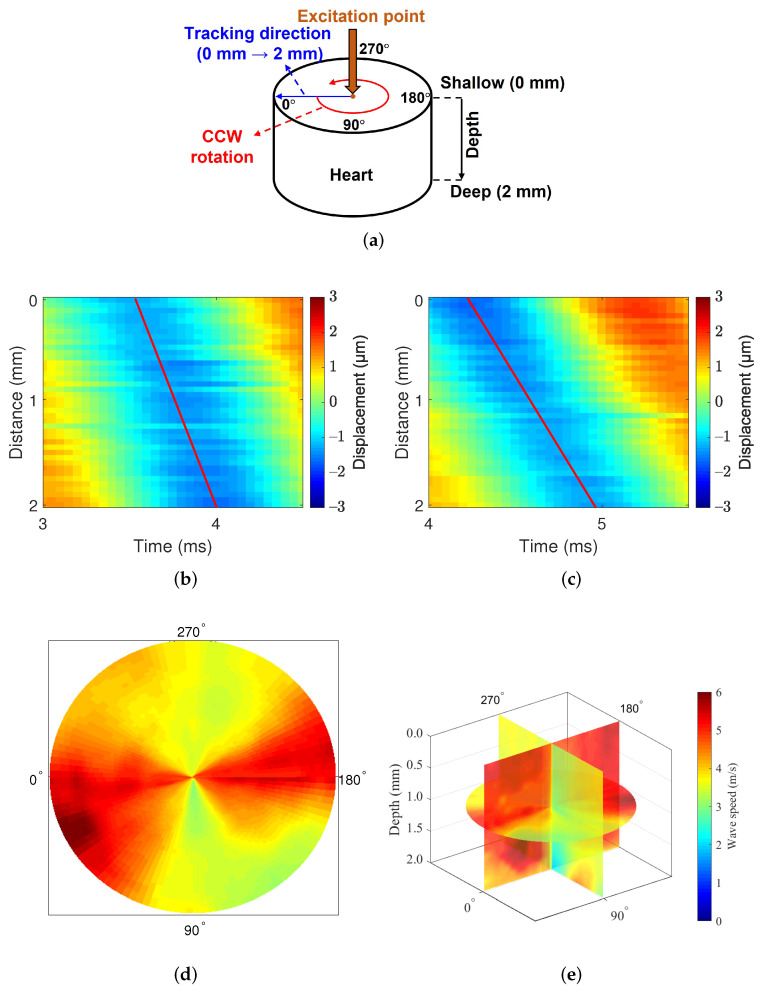
The use of imaging devices to assess directional mechanics of tissues is highly desirable. This is because the directional mechanics depend on fiber orientation, and altered directional mechanics are closely related to the pathological status of tissues. However, measuring directional mechanics in tissues with high-stiffness is challenging due to the difficulty of generating localized displacement in these tissues using acoustic radiation force, a general method for generating displacement in ultrasound-based elastography. In addition, common ultrasound probes do not provide rotational function, which makes the measurement of directional mechanics inaccurate and unreliable. Therefore, we developed a high-frequency ultrasound mechanical wave elastography system that can accommodate a wide range of tissue stiffness and is also equipped with a motorized rotation stage for precise imaging of directional mechanics. A mechanical shaker was applied to the elastography system to measure tissues with high-stiffness. Phantom and ex vivo experiments were performed. In the phantom experiments, the lateral and axial resolution of the system were determined to be 144 μm and 168 μm, respectively. In the ex vivo experiments, we used swine heart and cartilage, both of which are considered stiff. The elastography system allows us to acquire the directional mechanics with high angular resolution in the heart and cartilage. The results demonstrate that the developed elastography system is capable of imaging a wide range of tissues and has high angular resolution. Therefore, this system might be useful for the diagnostics of mechanically anisotropic tissues via ex vivo tests.
Keywords: cartilage experiment; elastography; heart experiment; high-frequency ultrasound; mechanical anisotropy; mechanical wave imaging.
Conflict of interest statement
The authors declare no conflict of interest.
Figures


Similar articles
-
Simultaneous measurement of tensile and shear elasticity and anisotropy in human skeletal muscle tissue using steered ultrasound shear waves.Acta Biomater. 2025 Jun 1;199:217-229. doi: 10.1016/j.actbio.2025.05.010. Epub 2025 May 3. Acta Biomater. 2025. PMID: 40324515
-
New Metric to Evaluate Cardiac Anisotropic Mechanics by Directional High-Frequency Ultrasound-Based Transverse Wave Elastography.IEEE Trans Ultrason Ferroelectr Freq Control. 2023 Jul;70(7):653-667. doi: 10.1109/TUFFC.2023.3279284. Epub 2023 Jun 29. IEEE Trans Ultrason Ferroelectr Freq Control. 2023. PMID: 37220030
-
Magnetic Resonance Elastography Reconstruction for Anisotropic Tissues.Med Image Anal. 2021 Dec;74:102212. doi: 10.1016/j.media.2021.102212. Epub 2021 Sep 20. Med Image Anal. 2021. PMID: 34587584
-
Deep learning in ultrasound elastography imaging: A review.Med Phys. 2022 Sep;49(9):5993-6018. doi: 10.1002/mp.15856. Epub 2022 Jul 30. Med Phys. 2022. PMID: 35842833 Review.
-
Advances in the clinical application of ultrasound elastography in uterine imaging.Insights Imaging. 2022 Sep 4;13(1):141. doi: 10.1186/s13244-022-01274-9. Insights Imaging. 2022. PMID: 36057675 Free PMC article. Review.
Cited by
-
Research progress of ultrasound in accurate evaluation of cartilage injury in osteoarthritis.Front Endocrinol (Lausanne). 2024 Aug 15;15:1420049. doi: 10.3389/fendo.2024.1420049. eCollection 2024. Front Endocrinol (Lausanne). 2024. PMID: 39211448 Free PMC article. Review.
References
-
- Lee W.N., Ingrassia C.M., Fung-Kee-Fung S.D., Costa K.D., Holmes J.W., Konofagou E.E. Theoretical quality assessment of myocardial elastography with in vivo validation. IEEE Trans. Ultrason. Ferroelectr. Freq. Control. 2007;54:2233–2245. - PubMed
MeSH terms
Grants and funding
LinkOut - more resources
Full Text Sources
