

Concentration of Microparticles Using Flexural Acoustic Wave in Sessile Droplets
- PMID: 35162014
- PMCID: PMC8839499
- DOI: 10.3390/s22031269
Concentration of Microparticles Using Flexural Acoustic Wave in Sessile Droplets
Abstract
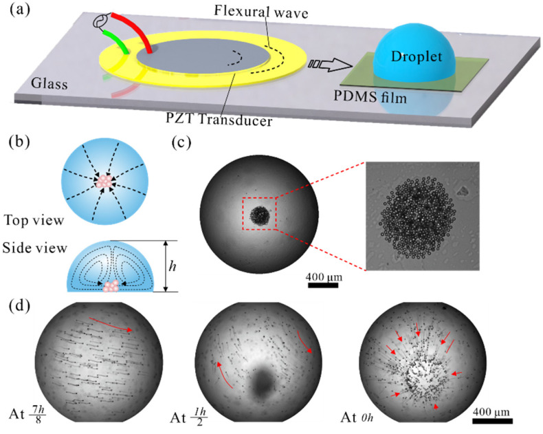
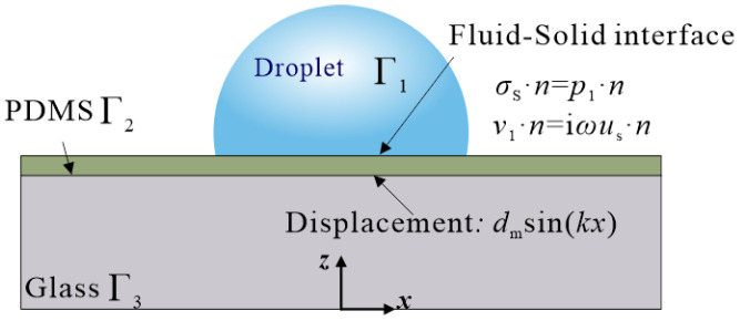
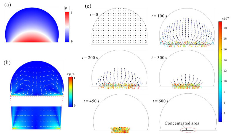
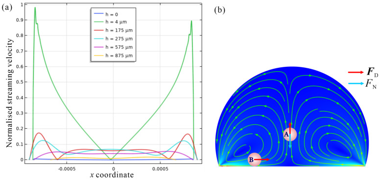
Acoustic manipulation of microparticles and cells has attracted growing interest in biomedical applications. In particular, the use of acoustic waves to concentrate particles plays an important role in enhancing the detection process by biosensors. Here, we demonstrated microparticle concentration within sessile droplets placed on the hydrophobic surface using the flexural wave. The design benefits from streaming flow induced by the Lamb wave propagated in the glass waveguide to manipulate particles in the droplets. Microparticles will be concentrated at the central area of the droplet adhesion plane based on the balance among the streaming drag force, gravity, and buoyancy at the operating frequency. We experimentally demonstrated the concentration of particles of various sizes and tumor cells. Using numerical simulation, we predicted the acoustic pressure and streaming flow pattern within the droplet and characterized the underlying physical mechanisms for particle motion. The design is more suitable for micron-sized particle preparation, and it can be valuable for various biological, chemical, and medical applications.
Keywords: acoustofluidic; flexural wave; numerical simulation; particle concentration.
Conflict of interest statement
The authors declare there is no conflict of interest.
Figures


Similar articles
-
Effect of microchannel protrusion on the bulk acoustic wave-induced acoustofluidics: numerical investigation.Biomed Microdevices. 2021 Dec 29;24(1):7. doi: 10.1007/s10544-021-00608-6. Biomed Microdevices. 2021. PMID: 34964071
-
Flexural wave-based soft attractor walls for trapping microparticles and cells.Lab Chip. 2021 Feb 9;21(3):582-596. doi: 10.1039/d0lc00865f. Lab Chip. 2021. PMID: 33355319 Free PMC article.
-
Low-frequency flexural wave based microparticle manipulation.Lab Chip. 2020 Apr 7;20(7):1281-1289. doi: 10.1039/d0lc00072h. Epub 2020 Mar 10. Lab Chip. 2020. PMID: 32154525 Free PMC article.
-
A simplified three-dimensional numerical simulation approach for surface acoustic wave tweezers.Ultrasonics. 2022 Sep;125:106797. doi: 10.1016/j.ultras.2022.106797. Epub 2022 Jun 30. Ultrasonics. 2022. PMID: 35780714 Review.
-
Acoustic Radiation Force: A Review of Four Mechanisms for Biomedical Applications.IEEE Trans Ultrason Ferroelectr Freq Control. 2021 Nov;68(11):3261-3269. doi: 10.1109/TUFFC.2021.3112505. Epub 2021 Oct 22. IEEE Trans Ultrason Ferroelectr Freq Control. 2021. PMID: 34520353 Review.
Cited by
-
Particle Manipulation in 2D Space Using a Capacitive Micromachined Ultrasonic Transducer.Micromachines (Basel). 2022 Mar 29;13(4):534. doi: 10.3390/mi13040534. Micromachines (Basel). 2022. PMID: 35457839 Free PMC article.
-
Recent progress in field-directed assembly of colloids in an evaporating droplet.Mater Today Bio. 2025 Jul 8;33:102072. doi: 10.1016/j.mtbio.2025.102072. eCollection 2025 Aug. Mater Today Bio. 2025. PMID: 40851655 Free PMC article. Review.
-
Cavity-agnostic acoustofluidic manipulations enabled by guided flexural waves on a membrane acoustic waveguide actuator.Microsyst Nanoeng. 2024 Mar 8;10:33. doi: 10.1038/s41378-023-00643-8. eCollection 2024. Microsyst Nanoeng. 2024. PMID: 38463549 Free PMC article.
References
MeSH terms
Grants and funding
LinkOut - more resources
Full Text Sources
