Review
. 2022 Jan 18;23(3):1014.
doi: 10.3390/ijms23031014.

The Role of Vitamin A in Retinal Diseases

Affiliations
Review

The Role of Vitamin A in Retinal Diseases

Jana Sajovic et al. Int J Mol Sci. .

Abstract

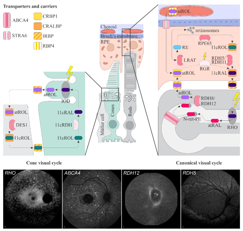
Vitamin A is an essential fat-soluble vitamin that occurs in various chemical forms. It is essential for several physiological processes. Either hyper- or hypovitaminosis can be harmful. One of the most important vitamin A functions is its involvement in visual phototransduction, where it serves as the crucial part of photopigment, the first molecule in the process of transforming photons of light into electrical signals. In this process, large quantities of vitamin A in the form of 11-cis-retinal are being isomerized to all-trans-retinal and then quickly recycled back to 11-cis-retinal. Complex machinery of transporters and enzymes is involved in this process (i.e., the visual cycle). Any fault in the machinery may not only reduce the efficiency of visual detection but also cause the accumulation of toxic chemicals in the retina. This review provides a comprehensive overview of diseases that are directly or indirectly connected with vitamin A pathways in the retina. It includes the pathophysiological background and clinical presentation of each disease and summarizes the already existing therapeutic and prospective interventions.

Keywords: ABCA4; RDH12; RDH5; RHO; retinal diseases; treatment; visual cycle; vitamin A.


Conflict of interest statement

The authors declare no conflict of interest.

Figures

Figure 1
Figure 1
Schematic representation of the canonical and cone visual cycles. Key enzymes, transporters, carriers and retinoids are shown. Fundus autofluorescence (FAF) images of representative patients, harbouring pathogenic variants in genes, encoding four essential proteins involved in the visual cycle, are presented below. Scale bars for four FAF images: 200 µm. Abbreviation expanation: RPE–retinal pigment epithelium, RBP4—retinol-binding protein 4, STRA6—stimulated by retinoic acid 6, CRBP1—cellular retinol-binding protein 1, LRAT—retinol:lecithin acyltransferase, RPE65—RPE-specific 65 kDa protein, RDH5—11-cis-retinol dehydrogenase 5, RDH11—11-cis-retinol dehydrogenase 11, CRALBP—cellular retinaldehyde-binding protein, RGR—retinal G protein-coupled receptor, IRBP—interphotoreceptor retinoid-binding protein, RDH8—all-trans-retinol dehydrogenases 8, RDH12—all-trans-retinol dehydrogenases 12, ABCA4—ATP-binding cassette subfamily A member 4, DES1—dihydroceramide desaturase-1, 11cRDH—11-cis-retinol dehydrogenase, RHO—rhodopsin, IOD—iodopsin, 11cRAL—11-cis-retinal, 11cROL—11-cis-retinol, atRAL—all-trans-retinal, atROL—all-trans-retinol, RE—retinyl esters, N-ret-PE—N-retinylidene-phosphatidylethanolamine, PE—phosphatidylethanolamine, A2E—N-retinyl-N-retinylidene ethanolamine. Source: Eye Hospital, University Medical Centre Ljubljana.
Figure 2
Figure 2
(A) Colour fundus image, (B) fundus infrared image and (C) FAF image in a patient with vitamin A deficiency. Note the corresponding (C1, arrow) spectral-domain optical coherence tomography (SD-OCT) image with abnormalities in photoreceptor outer segments, which (D, arrow) normalized after treatment with vitamin A supplementation. Patient’s best corrected Snellen decimal visual acuity before treatment was 0.7 on the right eye and 0.6 on the left eye. After treatment visual acuity improved to 1.0 on the right eye and 0.9 on the left eye. Scale bars: 200 µm. Source: Eye Hospital, University Medical Centre Ljubljana.
Figure 3
Figure 3
Clinical findings in a patient harbouring p.(Trp431*) and p.(Asp262Gly) in ABCA4. Mac-ular affection, fundus flecks and peripapillary sparring (diagnostic triad) are shown on (A) colour fundus image, (B) fundus infrared image and (C) FAF image. On corresponding (C1,C2) SD-OCT images, (arrowheads) hyperautofluorescent flecks, (arrow) RPE atrophy and (rectangle) peripapillary sparing can also be observed. Patient’s best corrected Snellen decimal visual acuity was 0.1 on both eyes. Scale bars: 200 µm. Source: Eye Hospital, University Medical Centre Ljubljana.
Figure 4
Figure 4
(A) Colour fundus image, (B) fundus infrared image and (C) FAF image with corresponding (C1) SD-OCT image showing clinical findings in a patient with p.(Gly90Asp) in RHO. Area within the arrowheads on C and C1 images corresponds to the preserved part of the retina. Patient’s best corrected Snellen decimal visual acuity was 0.6 on the right eye and 0.4 on the left eye. Scale bars: 200 µm. Source: Eye Hospital, University Medical Centre Ljubljana.
Figure 5
Figure 5
Clinical characteristics of a patient homozygous for p.(Thr137Ser) in RDH5. (A) Colour fundus image, (B) infrared image and (C) FAF image with corresponding (C1) SD-OCT image showing (arrowheads) retinal flecks. FAF image shows reduced autofluorescence in the entire retina. Patient’s best corrected Snellen decimal visual acuity was 0.8 on both eyes. Scale bars: 200 µm. Source: Eye Hospital, University Medical Centre Ljubljana.
Figure 6
Figure 6
(A,C) Ultra-widefield and 50° colour fundus images, (D) fundus infrared image, (B,E) ul-tra-widefield and 55° FAF images and (E1) SD-OCT image of a patient homozygous for p.(Ala126Glu) in RDH12. (A,C) Colour fundus images and (D) fundus infrared image demonstrate bone-spicule pigmentation and wide-spread retinal atrophy. The latest is also shown on (E1) OCT, which shows loss of the photoreceptor inner segment ellipsoid band and (arrow) atrophy of the RPE. Peripapillary retinal preservation can be seen on A, B, C, D and E images. The quality of E and E1 images is low due to reduced visual acuity and nystagmus. Patient’s best corrected Snellen decimal visual acuity was 0.3 on both eyes. Scale bars A, B: 2 mm; C, D, E and E1: 200 µm. Source: Eye Hospital, University Medical Centre Ljubljana.
Figure 7
Figure 7
Patient with Sorsby dystrophy due to pathogenic variant p.(Ser170Cys) in TIMP3. (A) Colour fundus image, (B) fundus infrared image, and (C) FAF image with corresponding (C1,C2) SD-OCT images showing (arrows) geographic atrophy and (arrowheads) subretinal drusen-like deposits. (D, arrow) Hyperfluorescent area is compatible with choroidal neovascularisation. (E) The reduced indocyanine green late-phase angiography fluorescence due to decreased permeability of Bruch’s membrane. Patient’s best corrected Snellen decimal visual acuity was 0.4 on the right eye and 0.6 on the left eye. Scale bars: 200 µm. Source: Eye Hospital, University Medical Centre Ljubljana.
Figure 8
Figure 8
Patient with age related macular degeneration. Representative (A) colour fundus image, (B) fundus infrared image and (C) FAF image with corresponding (C1) SD-OCT image show (arrowheads) reticular drusen. Patient’s best corrected Snellen decimal visual acuity was 0.8 on both eyes. Scale bars: 200 µm. Source: Eye Hospital, University Medical Centre Ljubljana.

References

    1. Carazo A., Macáková K., Matoušová K., Krčmová L.K., Protti M., Mladěnka P. Vitamin A Update: Forms, Sources, Kinetics, Detection, Function, Deficiency, Therapeutic Use and Toxicity. Nutrients. 2021;13:1703. doi: 10.3390/nu13051703. - DOI - PMC - PubMed
    1. Blaner W.S., Li Y., Brun P.J., Yuen J.J., Lee S.A., Clugston R.D. Vitamin A Absorption, Storage and Mobilization. Subcell Biochem. 2016;81:95–125. doi: 10.1007/978-94-024-0945-1_4. - DOI - PubMed
    1. O’Byrne S.M., Wongsiriroj N., Libien J., Vogel S., Goldberg I.J., Baehr W., Palczewski K., Blaner W.S. Retinoid absorption and storage is impaired in mice lacking lecithin:retinol acyltransferase (LRAT) J. Biol. Chem. 2005;280:35647–35657. doi: 10.1074/jbc.M507924200. - DOI - PMC - PubMed
    1. Orban T., Palczewska G., Palczewski K. Retinyl ester storage particles (retinosomes) from the retinal pigmented epithelium resemble lipid droplets in other tissues. J. Biol. Chem. 2011;286:17248–17258. doi: 10.1074/jbc.M110.195198. - DOI - PMC - PubMed
    1. Frey S.K., Vogel S. Vitamin A metabolism and adipose tissue biology. Nutrients. 2011;3:27–39. doi: 10.3390/nu3010027. - DOI - PMC - PubMed