

Active Materials for 3D Printing in Small Animals: Current Modalities and Future Directions for Orthopedic Applications
- PMID: 35162968
- PMCID: PMC8834768
- DOI: 10.3390/ijms23031045
Active Materials for 3D Printing in Small Animals: Current Modalities and Future Directions for Orthopedic Applications
Abstract
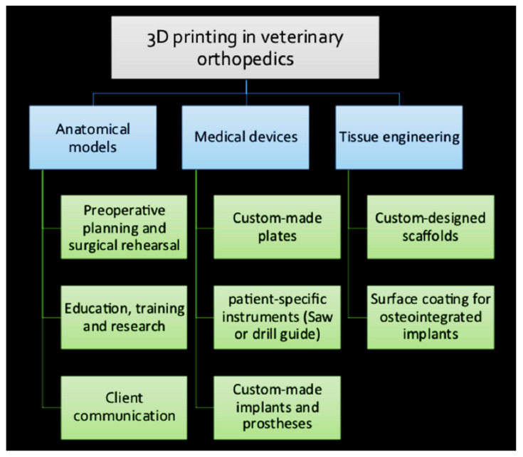
The successful clinical application of bone tissue engineering requires customized implants based on the receiver's bone anatomy and defect characteristics. Three-dimensional (3D) printing in small animal orthopedics has recently emerged as a valuable approach in fabricating individualized implants for receiver-specific needs. In veterinary medicine, because of the wide range of dimensions and anatomical variances, receiver-specific diagnosis and therapy are even more critical. The ability to generate 3D anatomical models and customize orthopedic instruments, implants, and scaffolds are advantages of 3D printing in small animal orthopedics. Furthermore, this technology provides veterinary medicine with a powerful tool that improves performance, precision, and cost-effectiveness. Nonetheless, the individualized 3D-printed implants have benefited several complex orthopedic procedures in small animals, including joint replacement surgeries, critical size bone defects, tibial tuberosity advancement, patellar groove replacement, limb-sparing surgeries, and other complex orthopedic procedures. The main purpose of this review is to discuss the application of 3D printing in small animal orthopedics based on already published papers as well as the techniques and materials used to fabricate 3D-printed objects. Finally, the advantages, current limitations, and future directions of 3D printing in small animal orthopedics have been addressed.
Keywords: 3D printing; materials; orthopedics; receiver-specific; veterinary.
Conflict of interest statement
The authors declare no conflict of interest.
Figures


Similar articles
-
3D-printed patient-specific applications in orthopedics.Orthop Res Rev. 2016 Oct 14;8:57-66. doi: 10.2147/ORR.S99614. eCollection 2016. Orthop Res Rev. 2016. PMID: 30774470 Free PMC article. Review.
-
3D printing metal implants in orthopedic surgery: Methods, applications and future prospects.J Orthop Translat. 2023 Sep 1;42:94-112. doi: 10.1016/j.jot.2023.08.004. eCollection 2023 Sep. J Orthop Translat. 2023. PMID: 37675040 Free PMC article. Review.
-
Patient-Specific 3-Dimensional-Printed Orthopedic Implants and Surgical Devices Are Potential Alternatives to Conventional Technology But Require Additional Characterization.Clin Orthop Surg. 2025 Feb;17(1):1-15. doi: 10.4055/cios23294. Epub 2024 Jun 26. Clin Orthop Surg. 2025. PMID: 39912074 Free PMC article. Review.
-
Application and Development of Modern 3D Printing Technology in the Field of Orthopedics.Biomed Res Int. 2022 Feb 15;2022:8759060. doi: 10.1155/2022/8759060. eCollection 2022. Biomed Res Int. 2022. PMID: 35211626 Free PMC article. Review.
-
Preoperative Planning Using 3D Printing Technology in Orthopedic Surgery.Biomed Res Int. 2021 Oct 12;2021:7940242. doi: 10.1155/2021/7940242. eCollection 2021. Biomed Res Int. 2021. PMID: 34676264 Free PMC article. Review.
Cited by
-
Patient-Specific 3D-Printed Osteotomy Guides and Titanium Plates for Distal Femoral Deformities in Dogs with Lateral Patellar Luxation.Animals (Basel). 2024 Mar 19;14(6):951. doi: 10.3390/ani14060951. Animals (Basel). 2024. PMID: 38540049 Free PMC article.
-
Apple Derived Exosomes Improve Collagen Type I Production and Decrease MMPs during Aging of the Skin through Downregulation of the NF-κB Pathway as Mode of Action.Cells. 2022 Dec 7;11(24):3950. doi: 10.3390/cells11243950. Cells. 2022. PMID: 36552714 Free PMC article.
-
3D printed orthopedic prostheses for domestic and wild birds-case reports.Sci Rep. 2024 Apr 5;14(1):7989. doi: 10.1038/s41598-024-58762-9. Sci Rep. 2024. PMID: 38580783 Free PMC article.
-
Evaluation of Patellar Groove Prostheses in Veterinary Medicine: Review of Technological Advances, Technical Aspects, and Quality Standards.Materials (Basel). 2025 Apr 3;18(7):1652. doi: 10.3390/ma18071652. Materials (Basel). 2025. PMID: 40271891 Free PMC article. Review.
-
Orthopedic applications of 3D printing in canine veterinary medicine.Front Vet Sci. 2025 Apr 23;12:1582720. doi: 10.3389/fvets.2025.1582720. eCollection 2025. Front Vet Sci. 2025. PMID: 40336818 Free PMC article.
References
-
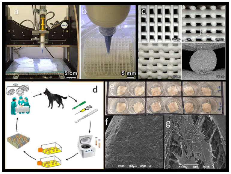
- Choi S., Oh Y.I., Park K.H., Lee J.S., Shim J.H., Kang B.J. New clinical application of three-dimensional-printed polycaprolactone/β-tricalcium phosphate scaffold as an alternative to allograft bone for limb-sparing surgery in a dog with distal radial osteosarcoma. J. Vet. Med. Sci. 2019;81:434–439. doi: 10.1292/jvms.18-0158. - DOI - PMC - PubMed
Publication types
MeSH terms
LinkOut - more resources
Full Text Sources
