. 2022 Jan 19;23(3):1083.
doi: 10.3390/ijms23031083.

Reduction in Ventilation-Induced Diaphragmatic Mitochondrial Injury through Hypoxia-Inducible Factor 1α in a Murine Endotoxemia Model

Affiliations

Reduction in Ventilation-Induced Diaphragmatic Mitochondrial Injury through Hypoxia-Inducible Factor 1α in a Murine Endotoxemia Model

Li-Fu Li et al. Int J Mol Sci. .

Abstract

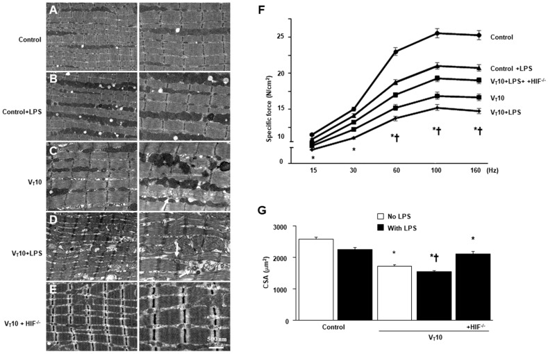
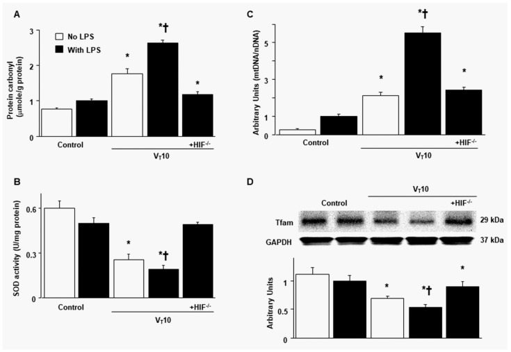
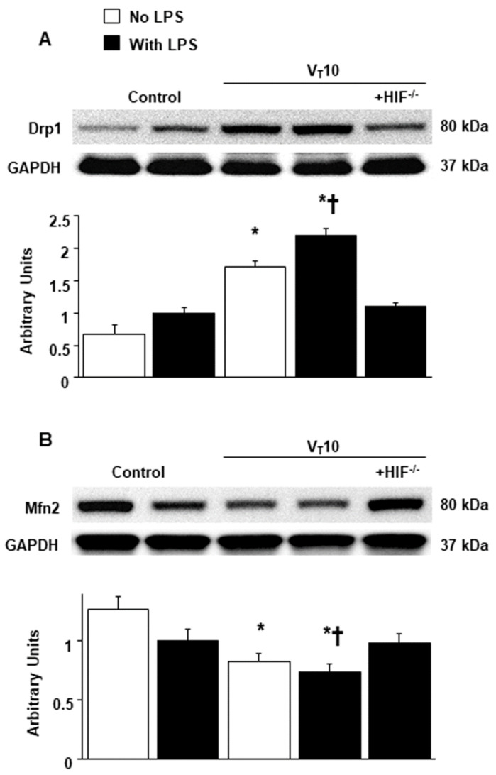
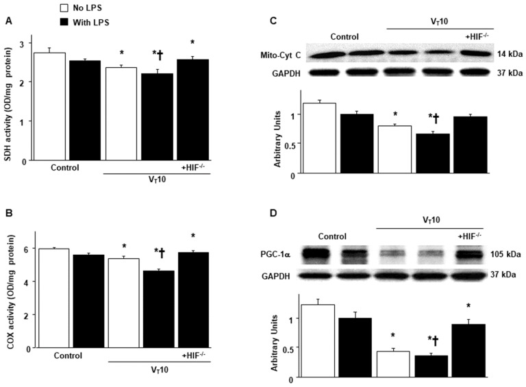
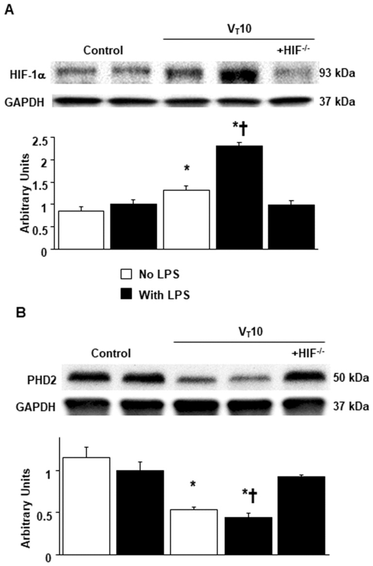
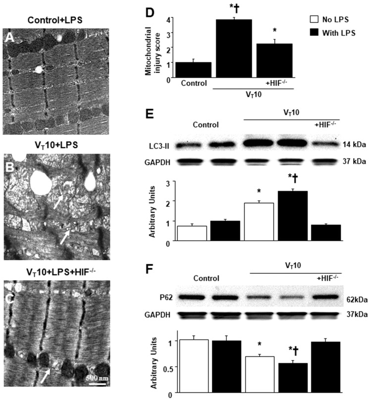
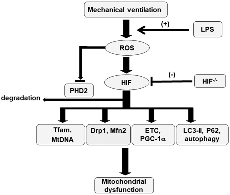
Mechanical ventilation (MV) is essential for patients with sepsis-related respiratory failure but can cause ventilator-induced diaphragm dysfunction (VIDD), which involves diaphragmatic myofiber atrophy and contractile inactivity. Mitochondrial DNA, oxidative stress, mitochondrial dynamics, and biogenesis are associated with VIDD. Hypoxia-inducible factor 1α (HIF-1α) is crucial in the modulation of diaphragm immune responses. The mechanism through which HIF-1α and mitochondria affect sepsis-related diaphragm injury is unknown. We hypothesized that MV with or without endotoxin administration would aggravate diaphragmatic and mitochondrial injuries through HIF-1α. C57BL/6 mice, either wild-type or HIF-1α-deficient, were exposed to MV with or without endotoxemia for 8 h. MV with endotoxemia augmented VIDD and mitochondrial damage, which presented as increased oxidative loads, dynamin-related protein 1 level, mitochondrial DNA level, and the expressions of HIF-1α and light chain 3-II. Furthermore, disarrayed myofibrils; disorganized mitochondria; increased autophagosome numbers; and substantially decreased diaphragm contractility, electron transport chain activities, mitofusin 2, mitochondrial transcription factor A, peroxisome proliferator activated receptor-γ coactivator-1α, and prolyl hydroxylase domain 2 were observed (p < 0.05). Endotoxin-stimulated VIDD and mitochondrial injuries were alleviated in HIF-1α-deficient mice (p < 0.05). Our data revealed that endotoxin aggravated MV-induced diaphragmatic dysfunction and mitochondrial damages, partially through the HIF-1α signaling pathway.

Keywords: autophagy; biogenesis; dynamics; hypoxia-inducible factor-1α; mitochondria; ventilator-induced diaphragm dysfunction.


Conflict of interest statement

The authors declare no conflict of interest.

Figures

Figure 1
Figure 1
Suppression of endotoxin-enhanced mechanical ventilation-induced diaphragm and mitochondrial injuries in HIF-1α-deficient mice. Representative micrographs of the longitudinal sections of diaphragm (×20,000: left panel; ×40,000: right panel) were from the same diaphragms of nonventilated control mice and mice ventilated at a tidal volume (VT) of 10 mL/kg (VT 10) for 8 h (n = 3 per group). (A,B) Nonventilated control wild-type mice with or without LPS treatment: normal sarcomeres with clear A bands, I bands, and Z bands; (C) 10 mL/kg wild-type mice without LPS treatment (normal saline): increase of diaphragmatic disarray; (D) 10 mL/kg wild-type mice with LPS treatment: disruption of sarcomeric structure, mitochondrial swelling with a vacuole-like structure, streaming of Z bands, and collection of lipid droplets; (E) 10 mL/kg HIF-1α deficient mice: reduction of diaphragmatic disruption. (F) Diaphragm muscle-specific force production was measured as described in Methods. (G) Cross-sectional area of diaphragm muscle fiber was measured as described in Methods (n = 5 per group). Mitochondrial swelling with concurrent formation of vacuoles and autophagosomes containing heterogeneous cargo are identified by arrows. * p < 0.05 versus the nonventilated control mice with LPS treatment; † p < 0.05 versus all other groups. Scale bar represents 500 nm. CSA = cross-sectional area; HIF−/− = hypoxia-inducible factor-1α-deficient mice; Hz = hertz; LPS = lipopolysaccharide; N = newton.
Figure 2
Figure 2
Inhibition of endotoxin-augmented mechanical ventilation-induced oxidative load, mitochondrial DNA, and mitochondrial transcription factor A in HIF-1α-deficient mice. (A) Protein carbonyl groups and (B) SOD were from the diaphragms of nonventilated control mice and mice ventilated at a tidal volume of 10 mL/kg for 8 h with or without LPS administration (n = 5 per group). (C) Real-time PCR performed for mitochondrial DNA expression was from the diaphragms of nonventilated control mice and mice ventilated at a tidal volume of 10 mL/kg for 8 h with or without LPS administration (n = 5 per group). Arbitrary units were expressed as the ratio of mitochondrial DNA to nuclear DNA (n = 5 per group). (D) Western blots were performed using antibodies that recognize mitochondrial transcription factor A and GAPDH expression from the diaphragms of nonventilated control mice and mice ventilated at a tidal volume of 10 mL/kg for 8 h with or without LPS administration (n = 5 per group). Arbitrary units were expressed as relative Tfam activation (n = 5 per group). * p < 0.05 versus the nonventilated control mice with LPS treatment; p < 0.05 versus all other groups. GAPDH = glyceraldehydes-phosphate dehydrogenase; mt = mitochondria; nDNA = nuclear DNA; SOD = superoxide dismutase; Tfam = mitochondrial transcription factor A.
Figure 3
Figure 3
Reduction of endotoxin-aggravated mechanical ventilation-mediated mitochondrial dynamics in HIF-1α-deficient mice. Western blots were performed using antibodies that recognize Drp1 (A), Mfn2 (B), and GAPDH expression from the diaphragms of nonventilated control mice and mice ventilated at a tidal volume of 10 mL/kg for 8 h with or without LPS administration (n = 5 per group). Arbitrary units were expressed as relative Drp1 and Mfn2 activation (n = 5 per group). * p < 0.05 versus the nonventilated control mice with LPS treatment; p < 0.05 versus all other groups. Drp1 = dynamin-related protein 1; Mfn2 = mitofusion 2.
Figure 4
Figure 4
Abrogation of endotoxin-enhanced mechanical ventilation-mediated mitochondrial biogenesis in HIF-1α-deficient mice. (A) SDH (complex II)) and (B) COX (complex IV) were from the diaphragms of nonventilated control mice and mice ventilated at a tidal volume of 10 mL/kg for 8 h with or without LPS administration (n = 5 per group). Western blots were performed using antibodies that recognize mitochondrial cytochrome C (C), PGC-1α (D), and GAPDH expression from the diaphragms of nonventilated control mice and mice ventilated at a tidal volume of 10 mL/kg for 8 h with or without LPS administration (n = 5 per group). Arbitrary units were expressed as relative mitochondrial cytochrome C and PGC-1α activation (n = 5 per group). * p < 0.05 versus the nonventilated control mice with LPS treatment; p < 0.05 versus all other groups. COX = cytochrome-c oxidase; Mito-Cyt C = mitochondrial cytochrome C; PGC-1α = peroxisome proliferator activated receptor-γ coactivator; SDH = succinate dehydrogenase.
Figure 5
Figure 5
Amelioration of endotoxin-augmented mechanical ventilation-mediated HIF-1α and PHD2 protein expression in HIF-1α-deficient mice. Western blots were performed using antibodies that recognize HIF-1α (A), PHD2 (B), and GAPDH expression from the diaphragms of nonventilated control mice and mice ventilated at a tidal volume of 10 mL/kg for 8 h with or without LPS administration (n = 5 per group). Arbitrary units were expressed as relative HIF-1α and PHD2 activation (n = 5 per group). * p < 0.05 versus the nonventilated control mice with LPS treatment; p < 0.05 versus all other groups. PHD2 = prolyl hydroxylase domain 2.
Figure 6
Figure 6
Inhibition of endotoxin-aggravated mechanical ventilation-induced diaphragm injury and autophagy in HIF-1α deficient mice. (AD) Representative micrographs of the longitudinal sections of diaphragm (×40,000) were from the diaphragms of nonventilated control mice and mice ventilated at a tidal volume of 10 mL/kg for 8 h with LPS administration (n = 3 per group). Mitochondrial damage with coexisting autophagosomes containing heterogeneous cargo, loss of cristae, and vacuole formation is identified by arrows. Western blots were performed using antibodies that recognize LC3-II (E), P62 (F), and GAPDH expression from the diaphragms of nonventilated control mice and mice ventilated at a tidal volume of 10 mL/kg for 8 h with or without LPS administration (n = 5 per group). Arbitrary units were expressed as relative LC3-II and P62 activation (n = 5 per group). * p < 0.05 versus the nonventilated control mice with LPS treatment; p < 0.05 versus all other groups. LC3-II = light chain 3-II.
Figure 7
Figure 7
Schematic figure illustrating the signaling pathway activation with mechanical ventilation and endotoxemia. Endotoxin-induced augmentation of mechanical stretch-mediated oxidative stress, mitochondrial dynamics and biogenesis, and autophagy, and diaphragm damage were alleviated with HIF-1α homozygous knockout. Drp1 = dynamin-related protein 1; ETC = electron transport chain; HIF = hypoxia-inducible factor; LC3-II = light chain 3-II; LPS = lipopolysaccharide; Mfn2 = mitofusion 2; Mt = mitochondria; PGC-1α = peroxisome proliferator activated receptor-γ coactivator-1α; PHD2 = prolyl hydroxylase domain 2; ROS = reactive oxygen species; Tfam = mitochondrial transcription factor A; VIDD = ventilator-induced diaphragm dysfunction.

References

    1. Horn A.G., Davis R.T., III., Baumfalk D.R., Kunkel O.N., Bruells C.S., McCullough D.J., Opoku-Acheampong A.B., Poole D.C., Behnke B.J. Impaired diaphragm resistance vessel vasodilation with prolonged mechanical ventilation. J. Appl. Physiol. 2019;127:423–431. doi: 10.1152/japplphysiol.00189.2019. - DOI - PMC - PubMed
    1. Pisani D.F., Dechesne C.A. Skeletal muscle HIF-1alpha expression is dependent on muscle fiber type. J. Gen. Physiol. 2005;126:173–178. doi: 10.1085/jgp.200509265. - DOI - PMC - PubMed
    1. Nguyen T.H., Conotte S., Belayew A., Decleves A.E., Legrand A., Tassin A. Hypoxia and Hypoxia-Inducible Factor Signaling in Muscular Dystrophies: Cause and Consequences. Int. J. Mol. Sci. 2021;22:7220. doi: 10.3390/ijms22137220. - DOI - PMC - PubMed
    1. Moarbes V., Mayaki D., Huck L., Leblanc P., Vassilakopoulos T., Petrof B.J., Hussain S.N.A. Differential regulation of myofibrillar proteins in skeletal muscles of septic mice. Physiol. Rep. 2019;7:e14248. doi: 10.14814/phy2.14248. - DOI - PMC - PubMed
    1. Azuelos I., Jung B., Picard M., Liang F., Li T., Lemaire C., Giordano C., Hussain S., Petrof B.J. Relationship between Autophagy and Ventilator-induced Diaphragmatic Dysfunction. Anesthesiology. 2015;122:1349–1361. doi: 10.1097/ALN.0000000000000656. - DOI - PubMed

MeSH terms

Substances