. 2022 Jan 20;23(3):1094.
doi: 10.3390/ijms23031094.

Detecting Bacterial-Human Lateral Gene Transfer in Chronic Lymphocytic Leukemia

Affiliations

Detecting Bacterial-Human Lateral Gene Transfer in Chronic Lymphocytic Leukemia

Ekaterina Akimova et al. Int J Mol Sci. .

Abstract

Chronic lymphocytic leukemia (CLL) is a very common and mostly incurable B-cell malignancy. Recent studies revealed high interpatient mutational heterogeneity and worsened therapy response and survival of patients with complex genomic aberrations. In line with this, a better understanding of the underlying mechanisms of specific genetic aberrations would reveal new prognostic factors and possible therapeutic targets. It is known that chromosomal rearrangements including DNA insertions often play a role during carcinogenesis. Recently it was reported that bacteria (microbiome)-human lateral gene transfer occurs in somatic cells and is enriched in cancer samples. To further investigate this mechanism in CLL, we analyzed paired-end RNA sequencing data of 45 CLL patients and 9 healthy donors, in which we particularly searched for bacterial DNA integrations into the human somatic genome. Applying the Burrows-Wheeler aligner (BWA) first on a human genome and then on bacterial genome references, we differentiated between sequencing reads mapping to the human genome, to the microbiome or to bacterial integrations into the human genome. Our results indicate that CLL samples featured bacterial DNA integrations more frequently (approx. two-fold) compared to normal samples, which corroborates the latest findings in other cancer entities. Moreover, we determined common integration sites and recurrent integrated bacterial transcripts. Finally, we investigated the contribution of bacterial integrations to oncogenesis and disease progression.

Keywords: CLL; bacterial integrations; lateral gene transfer (LGT).


Conflict of interest statement

The authors declare no conflict of interest.

Figures

Figure 1
Figure 1
Detecting bacterial integrations into the human genome in CLL patients—analysis workflow. RNAseq data were aligned to the human genome (hg19) and subsequently to bacterial genome references via BWA. The reads supporting LGT were defined as reads featuring only one mate mapping exclusively to human genome and only one mate mapping exclusively to bacterial genome.
Figure 2
Figure 2
Reads supporting bacterial–human LGT were enriched in CLL versus non-leukemic B cells. (A) Distribution of reads supporting LGT in CLL samples (red) and in healthy B cells (green). The graph shows the percentage of samples featuring indicated number of reads supporting LGT. (B) The figure displays a comparison between CLL samples from CLL patients and B-cell samples from healthy (H) donors, whereas percentage of reads supporting bacterial–human lateral gene transfer is shown. Median is indicated. Significance was calculated using unpaired two-tailed t-test. Below, key information is summarized for both groups.
Figure 3
Figure 3
Genomic regions affected by bacterial–human LGT. (A) The pie chart shows the distribution of LGT events in the CLL cohort among different gene coding structures: most of them (36%) occurred in intronic regions. (B) The bar plot shows the top 30 genes affected by LGT. The Y-axis indicates the percentage of patients in each cohort (CLL in pink, healthy in blue) featuring a bacterial integration in the respective gene.
Figure 4
Figure 4
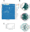
Distribution of bacterial species contributing to LGT in CLL. (A) The heat map shows the number of bacterial transcripts integrated into human genome from the 20 most abundant bacteria genera for each CLL sample. In total, we detected 216 different bacteria genera across the entire dataset. (B) Circos plots illustrate the integration sites of three selected bacteria genera (Pseudomonas sp., Mesorhizobium sp. and Acinetobacter sp.) into the human genome. (C) MDS analysis of LGT event counts, using a matrix that lists the number of LGT events for each bacteria genus per patient.
Figure 5
Figure 5
Correlation of LGT events with clinically relevant parameters. (A) Kaplan–Meier overall survival (OS, left) and progression-free survival (PFS, right) curves comparing patients grouped according to MDS analysis of LGT events (groups 1 and 2). (B) Kaplan–Meier overall survival (OS, left) and progression-free survival (PFS, right) curves comparing patients with higher (LGThigh, in yellow) and lower (LGTlow, in blue) amounts of bacterial integrations (cutoffs calculated by Cox regression analysis). For all survival plots’ p-values, confidence intervals as well as horizontal and vertical median survival lines are indicated. The stacked bar plots show the percentage of patients in group 1 and group 2 either featuring IGVH mutation (C), del11q (D), del17q (E), tri12 (F), del13q (G) or not. (H) The stacked bar plot indicates the percentage of patients in group 1 and group 2 with either high or low CD38. For (CH), significances were calculated with a two-sided Fisher’s exact test. (I) The stacked bar plot displays the percentage of patients in group 1 and group 2 in different stages according to the assigned RAI index. Significance was calculated with Pearson’s chi-squared test.

References

    1. Pleyer L., Egle A., Hartmann T.N., Greil R. Molecular and Cellular Mechanisms of CLL: Novel Therapeutic Approaches. Nat. Rev. Clin. Oncol. 2009;6:405–418. doi: 10.1038/nrclinonc.2009.72. - DOI - PubMed
    1. Landau D.A., Carter S.L., Stojanov P., McKenna A., Stevenson K., Lawrence M.S., Sougnez C., Stewart C., Sivachenko A., Wang L., et al. Evolution and Impact of Subclonal Mutations in Chronic Lymphocytic Leukemia. Cell. 2013;152:714–726. doi: 10.1016/j.cell.2013.01.019. - DOI - PMC - PubMed
    1. Landau D.A., Tausch E., Taylor-Weiner A.N., Stewart C., Reiter J.G., Bahlo J., Kluth S., Bozic I., Lawrence M., Böttcher S., et al. Mutations Driving CLL and Their Evolution in Progression and Relapse. Nature. 2015;526:525–530. doi: 10.1038/nature15395. - DOI - PMC - PubMed
    1. Chai-Adisaksopha C., Brown J.R. FCR Achieves Long-Term Durable Remissions in Patients with IGHV-Mutated CLL. Blood. 2017;130:2278–2282. doi: 10.1182/blood-2017-07-731588. - DOI - PubMed
    1. Visentin A., Facco M., Gurrieri C., Pagnin E., Martini V., Imbergamo S., Frezzato F., Trimarco V., Severin F., Raggi F., et al. Prognostic and Predictive Effect of IGHV Mutational Status and Load in Chronic Lymphocytic Leukemia: Focus on FCR and BR Treatments. Clin. Lymphoma Myeloma Leuk. 2019;19:678–685.e4. doi: 10.1016/j.clml.2019.03.002. - DOI - PubMed

MeSH terms

LinkOut - more resources