. 2022 Jan 20;23(3):1131.
doi: 10.3390/ijms23031131.

Rab21 Protein Is Degraded by Both the Ubiquitin-Proteasome Pathway and the Autophagy-Lysosome Pathway

Affiliations

Rab21 Protein Is Degraded by Both the Ubiquitin-Proteasome Pathway and the Autophagy-Lysosome Pathway

Pinduo Liu et al. Int J Mol Sci. .

Abstract

Rab21 is a GTPase protein that is functional in intracellular trafficking and involved in the pathologies of many diseases, such as Alzheimer's disease (AD), glioma, cancer, etc. Our previous work has reported its interaction with the catalytic subunit of gamma-secretase, PS1, and it regulates the activity of PS1 via transferring it from the early endosome to the late endosome/lysosome. However, it is still unknown how Rab21 protein itself is regulated. This work revealed that Rab21 protein, either endogenously or exogenously, can be degraded by the ubiquitin-proteasome pathway and the autophagy-lysosome pathway. It is further observed that the ubiquitinated Rab21 is increased, but the total protein is unchanged in AD model mice. We further observed that overexpression of Rab21 leads to increased expression of a series of genes involved in the autophagy-lysosome pathway. We speculated that even though the ubiquitinated Rab21 is increased due to the impaired proteasome function in the AD model, the autophagy-lysosome pathway functions in parallel to degrade Rab21 to keep its protein level in homeostasis. In conclusion, understanding the characters of Rab21 protein itself help explore its potential as a target for therapeutic strategy in diseases.

Keywords: Rab21 protein; autophagy-lysosome pathway; ubiquitin-proteasome pathway.


Conflict of interest statement

The authors declare that they have no competing interests.

Figures

Figure 1
Figure 1
The time-pulse chase assay for the half-life and the proteasome/lysosome-mediated degradation of Rab21 protein. (A) Rab21 plasmid (pcDNA4-Rab21) was transfected into HEK293 cells using Lipofectamine 2000. The transfected cells were treated with 100 μmol/L cycloheximide for 0, 8, 16, 20, 24, 28 h and then harvested. Rab21 proteins were detected by Western blot. (B) The protein level of Rab21 was shown by a line chart. (C) Rab21 plasmid transfected HEK293 cells were treated with vehicle solution control or 10 μmol/L MG132 or 10 mmol/L ammonium chloride for 24 h. (D) Rab21 protein levels were significantly increased by proteasomal and lysosomal inhibitor treatment. Protein levels were normalized to β-actin controls. Data are presented as mean ± SD. N = 3. * p < 0.05 relative to control by ANOVA.
Figure 2
Figure 2
The proteasome- and lysosome-dependent proteolysis of exogenous Rab21. (A) Rab21 plasmid (pcDNA4-Rab21) was transfected into HEK293 cells using Lipofectamine 2000. The transfected cells were treated with 10 μmol/L lactacystin for 0–24 h for a time-course assay. (B) Rab21 plasmid transfected HEK293 cells were treated with vehicle or lactacystin with gradient concentrations (0, 1, 2, 5, 10, 20 μmol/L) for 24 h. Rab21 protein levels were gradually increased in a lactacystin time- (C) and dosage- (D) dependent manner. (E) Rab21-containing plasmid transfected HEK293 cells were treated with 10 mmol/L ammonium chloride for 0–24 h’s time-dependent assay. (F) Rab21 plasmid transfected Hek293 cells were treated with gradient concentration (0, 1, 5, 10, 20, 50 mmol/L) of ammonium chloride for 24 h. Rab21 protein levels gradually increased in ammonium chloride in a time- (G) and dose- (H) dependent manner. Data are presented as mean ± SD. N = 3. * p < 0.05, ** p < 0.01, *** p < 0.001 relative to control by ANOVA.
Figure 3
Figure 3
The proteasome- and lysosome-dependent proteolysis of endogenous Rab21 protein (A) SH-SY5Y cells were treated with 10 μmol/L lactacystin for 0–24 h time-course assay. (B) SH-SY5Y cells were treated with vehicle or lactacystin with gradient concentrations (0, 1, 2, 5, 10, 20 μmol/L) for 24 h. Rab21 protein levels in the lactacystin time- (C) and dosage- (D) dependent experiments were significantly increased. (E) Cells were treated with 10 mmol/L ammonium chloride for 0–24 h time-dependent assay. (F) Cells were treated with gradient concentrations (0, 1, 5, 10, 20, 50 mmol/L) of ammonium chloride for 24 h. Rab21 protein levels in the ammonium chloride time- (G) and dosage- (H) dependent experiments were significantly increased. Data are presented as mean ± SD. N = 3. * p < 0.05, ** p < 0.01, *** p < 0.001 relative to control by ANOVA.
Figure 4
Figure 4
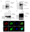
The proteasome-mediated degradation of Rab21 is ubiquitin-dependent. (A) Hek293 cells were transfected with Rab21 plasmid or pCDNA4 plasmid for 48 h. Cell lysates were immunoprecipitated with anti-ubiquitin followed by immunoblotting with anti-Rab21. (B) Hek293 cells were transfected with Rab21 plasmid or pCDNA4 plasmid for 48 h. Cell lysates were immunoprecipitated with anti-Rab21 followed by immunoblotting with anti-ubiquitin. (C,D) SH-SY5Y cells were treated with either vehicle or lactacystin or MG132 for 24 h. Cell lysates were immunoprecipitated with anti-Rab21 followed by immunoblotting with anti-ubiquitin. (E) SH-SY5Y cells were treated with either vehicle or lactacystin (10 μmol/L) for 24 h. Cells were fixed and incubated with primary antibody of Rab21 and ubiquitin, then incubated with Alexa Fluor® 488 (Abcam, Cambridge, UK) conjugated donkey anti-rabbit secondary antibody and Alexa Fluor® 555 (Abcam, Cambridge, UK) conjugated donkey anti-mouse secondary antibody. Proteasomal inhibitor treatment caused accumulation of ubiquitinated Rab21. Scale bar = 10 μm. Magnification: ×100.
Figure 5
Figure 5
The level of Rab21 and the ubiquitination of Rab21 in AD model mice (A) Rab21 mRNA levels in the brain of WT and 5 × FAD mice were detected by real-time PCR. Rab21 mRNA quantifications were normalized to β-actin. (B) Rab21 protein levels in the cerebral cortex and hippocampus of WT and 5 × FAD mice were examined by Western blot. (C) Lysates of the cerebral cortex were immunoprecipitated with anti-ubiquitin followed by immunoblotting with anti-Rab21. (D) Beclin1 protein level in the hippocampus of WT and 5 × FAD mice were examined by Western blot. Data presented as mean ± SD. N = 4 for WT and N = 4 for 5 × FAD mice (9 months old). Statistical analyses were performed using Student’s t-test. * p < 0.05 relative to control by ANOVA.
Figure 6
Figure 6
Rab21 upregulates genes involved in the autophagy-lysosome pathway. (A) SH-SY5Y cells were transfected with Rab21 plasmid for 48 h. Autophagy-lysosome pathway-related genes were detected by real-time PCR. (B) Cell lysates were immunoblotted with anti-LC3. LC3 II/I was significantly increased by overexpression of Rab21. (C) SH-SY5Y cells were transfected with Rab21 plasmid for 48 h. Cells were fixed and incubated with primary antibody of Rab21 and LC3, then incubated with Alexa Fluor® 488 conjugated donkey anti-rabbit secondary antibody and Alexa Fluor® 555 conjugated donkey anti-mouse secondary antibody. Scale bar = 10 μm. Magnification: ×100. Data are presented as mean ± SD. N = 3. * p < 0.05, ** p < 0.01, *** p < 0.001 relative to control by ANOVA.

Similar articles

Cited by

References

    1. Del Olmo T., Lacarrière-Keïta C., Normandin C., Jean D., Boisvert F.-M., Jean S. RAB21 interacts with TMED10 and modulates its localization and abundance. Biol. Open. 2019;8:bio045336. doi: 10.1242/bio.045336. - DOI - PMC - PubMed
    1. Vicinanza M., D’angelo G., Di Campli A., De Matteis M.A. Function and dysfunction of the PI system in membrane trafficking. EMBO J. 2008;27:2457–2470. doi: 10.1038/emboj.2008.169. - DOI - PMC - PubMed
    1. Pylypenko O., Hammich H., Yu I.-M., Houdusse A. Rab GTPases and their interacting protein partners: Structural insights into Rab functional diversity. Small GTPases. 2018;9:22–48. doi: 10.1080/21541248.2017.1336191. - DOI - PMC - PubMed
    1. Hutagalung A.H., Novick P.J. Role of Rab GTPases in membrane traffic and cell physiology. Physiol. Rev. 2011;91:119–149. doi: 10.1152/physrev.00059.2009. - DOI - PMC - PubMed
    1. Zhou F., Zou S., Chen Y., Lipatova Z., Sun D., Zhu X., Li R., Wu Z., You W., Cong X., et al. A Rab5 GTPase module is important for autophagosome closure. PLoS Genet. 2017;13:e1007020. doi: 10.1371/journal.pgen.1007020. - DOI - PMC - PubMed

LinkOut - more resources