. 2022 Jan 20;23(3):1135.
doi: 10.3390/ijms23031135.

Effect of Myosin Isoforms on Cardiac Muscle Twitch of Mice, Rats and Humans

Affiliations

Effect of Myosin Isoforms on Cardiac Muscle Twitch of Mice, Rats and Humans

Momcilo Prodanovic et al. Int J Mol Sci. .

Abstract

To understand how pathology-induced changes in contractile protein isoforms modulate cardiac muscle function, it is necessary to quantify the temporal-mechanical properties of contractions that occur under various conditions. Pathological responses are much easier to study in animal model systems than in humans, but extrapolation between species presents numerous challenges. Employing computational approaches can help elucidate relationships that are difficult to test experimentally by translating the observations from rats and mice, as model organisms, to the human heart. Here, we use the spatially explicit MUSICO platform to model twitch contractions from rodent and human trabeculae collected in a single laboratory. This approach allowed us to identify the variations in kinetic characteristics of α- and β-myosin isoforms across species and to quantify their effect on cardiac muscle contractile responses. The simulations showed how the twitch transient varied with the ratio of the two myosin isoforms. Particularly, the rate of tension rise was proportional to the fraction of α-myosin present, while the β-isoform dominated the rate of relaxation unless α-myosin was >50%. Moreover, both the myosin isoform and the Ca2+ transient contributed to the twitch tension transient, allowing two levels of regulation of twitch contraction.

Keywords: MUSICO platform; calcium sensitivity; cross-species simulation; level of incorporation; tension relaxation; trabeculae.


Conflict of interest statement

The authors declare no conflict of interest and this article reflects only the authors’ views. The European Commission is not responsible for any use that may be made of the information it contains.

Figures

Figure 1
Figure 1
Schematic representation of MUSICO platform in reference to the 3D half-sarcomere structure. (A) Kinetic scheme of calcium-binding to cTnC and interaction of cTnI with actin in cardiac muscle. Calcium-binding to cTnC forms a CacTnC complex, with an equilibrium rate of KCa=K˜Ca·[Ca2+], where K˜Ca is a rate constant and [Ca2+] is the instantaneous calcium concentration. Conformational changes within the cTn molecule forming the cTnC.cTnI or CacTnC.TnI complex, which have a low affinity to actin, are defined by the equilibrium state transition rate λ. In the CacTnC.cTnI (or cTnC.cTnI) state(s), cTnI is dissociated from actin, enabling Tpm to move freely, mostly azimuthally, and permitting myosin-binding and force generation. The rate of calcium-binding to cTnC.TnI to form a CacTnC.cTnI complex is accelerated by a factor of 1/εo. (B) Six-state model of the actomyosin cycle includes five biochemical states consistent with observed structural states: M.D.Pi, A.M.D.Pi, A.M.D, A.M and M.T. The strain-dependent state transition rates are associated with conformational changes defining the structural conformations of myosin in each state (for details, see [1,15]). An additional sixth state, the so-called “parked state” (PS), represents the interaction of myosin heads with the thick filament backbone. The transition rate from PS to M.D.Pi is assumed to be strongly dependent on [Ca2+], and the reverse rate is independent of [Ca2+]. (C) The 3D structural organization of sarcomeres includes thick (myosin) filaments interdigitated with thin filaments composed of actin and regulatory proteins, along with the ancillary protein titin. The 3D formulation includes an azimuthal shift of myosin crowns (Lc = 1, 2, 3), where the interaction of crossbridges with the target zones (green filled circles) on an actin filament is defined by axial crossbridge distortion and azimuthal angles defining the positions of actin filaments relative to the myosin filament and the positions of myosin-binding sites on actin (for details, see [21]). The parameters defining the lattice are the half-sarcomere length (HSL), the length of the actin filament (La) and the interfilament spacing (d10). The trabecula elasticity (SE) in series with the contractile element (CE) consisting of multiple sarcomere muscle fibers [1] is symbolically displayed here as the SE and 3D half-sarcomere structure, respectively. The thin filament sketch is adapted from the fragment in figure of Gordon et al. [23] and the thick filament from Anderson and Granzier [24].
Figure 2
Figure 2
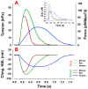
MUSICO predictions of twitch contractions in mouse, rat and human fixed-length trabeculae (A). The predicted tensions (thick solid lines) showed excellent agreement with those observed in mice, rat and human trabeculae (Chung et al. [34]) (thin dashed lines). The fits were achieved with a six-state crossbridge cycle that includes a parked state (PS) with calcium-dependent kPS and trabeculae series elasticity (SE). The crossbridge cycle state transition rates were for a completely α-cardiac myosin isoform in mice and a predominantly β-isoform in human simulations. In rats, the α- and β-isoform homodimers were randomly distributed, helically arranged myosin head pairs along a thick filament, containing an average of 75% α and 25% β. Following the experiments of Chung et al. [34], the simulations were done on fixed-length trabeculae at a relaxed sarcomere length of 2.2 μm and at 25 °C. The calcium transients (inset) were taken from rat right ventricle (RV) trabeculae at 25 °C (Janssen et al. [35]). The mouse calcium transient was derived from Ferrantini et al. [36] and the human calcium transients were taken from Ferrantini et al. [37]. The common model parameters are shown in Table 1 and parameters specific to mouse, rat and human trabeculae are shown in Table 2. MUSICO simulations also predicted a change in the half-sarcomere length, denoted as “Chng. HSL” in (B). The changes in the sarcomere length were not experimentally recorded in mice, rats or humans, but the predicted displacements from the rat simulations were similar to the observations of Caremani et al. [28] in rat fixed-length trabeculae.
Figure 3
Figure 3
Procedure for modeling twitch contractions of different species. Stage I: Based on the previously published mouse twitch, adjusted for the conditions of Chung et al. [34], and the data of Deacon et al. [8] for the crossbridge cycle parameters of mouse α- and human β-myosins. Stage II: Based on Stage 1 and the data of Deacon et al. [38] on human α-myosin, which were then extrapolated to mouse β-myosin. Stage III: Based on the previous publication on the rat twitch (Mijailovich et al. [1]) and interpolating the crossbridge values for rats between those of mice and humans. Stage IV: Using the crossbridge parameter values derived in Stages IIII to predict the effect of variable ratios of α- and β-myosin in each case.
Figure 4
Figure 4
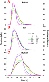
Effect of various fractions of α- and β-isoforms on twitch responses in fixed-end isometric conditions from mouse (A), rat (B) and human (C) trabeculae. The twitches show about the same twitch tension but the mouse (A) shows a much faster tension rise and tension relaxation than the rat (B). These parameters are much slower in the human for both the α- and β-isoforms (C), signifying differences in kinetics parameters of the same isoform across the species (Table 2). These responses were simulated assuming the same Ca2+ transient as in Figure 2. The differences between the three species, therefore, also reflect the differences in the magnitude of the peak and duration of respective Ca2+ transients.
Figure 5
Figure 5
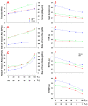
Effects of fractions of α- vs. β-isoforms on twitch characteristics. Parameters plotted are (A) peak tension (PT), (B) rates of tension rise and (C) relaxation, defined as a slope between 30 and 75% of the PT during the rise and vice versa during relaxation, (D) peak duration, defined as the time during which the tension is above 90% of the PT, (E) TTP, defined as the time from the onset of twitch contraction till the tension reaches the peak, (F) RT50% and (G) RT90%, defined as the time intervals from the time when the PT is reached to the times at which the tension relaxes to 50% and 90% of the PT, respectively. The magnitude of the peak tension and the rate of tension rise approximately linearly increased with the increase of α-isoform fraction, while the peak duration and TTP linearly decreased as the % α increased. The rate of tension relaxation nonlinearly increased while the RT50% and RT90% nonlinearly decreased.
Figure 6
Figure 6
Force-pCa relations of trabeculae containing (A) 100% α- or (B) 100% β-isoforms in mouse, rat and human cardiac muscles. At full activation at pCa < 5.5, all cases showed similar tension with differences between the species of <20 kPA or <20% of the largest tension, and even less between the corresponding α and β in the same species. For the α- and β-isoforms, the mouse showed the highest and the rat the lowest tension. Interestingly, all showed very similar Hill coefficient nH and pCa50 values.

Similar articles

Cited by

References

    1. Mijailovich S.M., Prodanovic M., Poggesi C., Geeves M.A., Regnier M. Multiscale modeling of twitch contractions in cardiac trabeculae. J. Gen. Physiol. 2021;153:e202012604. doi: 10.1085/jgp.202012604. - DOI - PMC - PubMed
    1. Cappelli V., Bottinelli R., Poggesi C., Moggio R., Reggiani C. Shortening velocity and myosin and myofibrillar ATPase activity related to myosin isoenzyme composition during postnatal development in rat myocardium. Circ. Res. 1989;65:446–457. doi: 10.1161/01.RES.65.2.446. - DOI - PubMed
    1. Chizzonite R.A., Zak R. Regulation of myosin isoenzyme composition in fetal and neonatal rat ventricle by endogenous thyroid hormones. J. Biol. Chem. 1984;259:12628–12632. doi: 10.1016/S0021-9258(18)90792-1. - DOI - PubMed
    1. Miyata S., Minobe W., Bristow M.R., Leinwand L.A. Myosin Heavy Chain Isoform Expression in the Failing and Nonfailing Human Heart. Circ. Res. 2000;86:386–390. doi: 10.1161/01.RES.86.4.386. - DOI - PubMed
    1. Reiser P.J., Portman M.A., Ning X.-H., Moravec C.S. Human cardiac myosin heavy chain isoforms in fetal and failing adult atria and ventricles. Am. J. Physiol. Circ. Physiol. 2001;280:H1814–H1820. doi: 10.1152/ajpheart.2001.280.4.H1814. - DOI - PubMed

LinkOut - more resources