Review
. 2022 Jan 20;23(3):1138.
doi: 10.3390/ijms23031138.

Retinal Plasticity

Affiliations
Review

Retinal Plasticity

Enrica Strettoi et al. Int J Mol Sci. .

Abstract

Brain plasticity is a well-established concept designating the ability of central nervous system (CNS) neurons to rearrange as a result of learning, when adapting to changeable environmental conditions or else while reacting to injurious factors. As a part of the CNS, the retina has been repeatedly probed for its possible ability to respond plastically to a variably altered environment or to pathological insults. However, numerous studies support the conclusion that the retina, outside the developmental stage, is endowed with only limited plasticity, exhibiting, instead, a remarkable ability to maintain a stable architectural and functional organization. Reviewed here are representative examples of hippocampal and cortical paradigms of plasticity and of retinal structural rearrangements found in organization and circuitry following altered developmental conditions or occurrence of genetic diseases leading to neuronal degeneration. The variable rate of plastic changes found in mammalian retinal neurons in different circumstances is discussed, focusing on structural plasticity. The likely adaptive value of maintaining a low level of plasticity in an organ subserving a sensory modality that is dominant for the human species and that requires elevated fidelity is discussed.

Keywords: deafferentation; remodeling; retinitis pigmentosa; structural plasticity.


Conflict of interest statement

The authors declare no conflict of interest. The funders had no role in the design of the study; the collection, analyses or interpretation of data; the writing of the manuscript; or in the decision to publish the results.

Figures

Figure 1
Figure 1
Rod bipolar cells (in green) of wild-type (A) and degenerating retinas (B), stained with antibodies against PKCα. Blue: nuclear counterstaining. Upon photoreceptor degeneration, the outer nuclear layer (ONL) becomes thinner and dendritic arbors of bipolar cells progressively retract from the outer plexiform layer (OPL) (arrows), while the complexity of their axonal arbors in the inner plexiform layer (IPL) decreases. These are typical changes occurring in neurons as a reaction to deafferentation. Abbreviations are the same in the following figures.
Figure 2
Figure 2
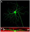
Example of retinal ganglion cell (A) from a Thy-1-GFP expressing mouse strain. The cell shows a narrow pattern of dendritic stratification in sublamina b (the innermost tier) of the IPL (A’). This pattern is not altered by the rd1 mutation hosted by this strain, one of the most aggressive genetic defects, leading to a rapid degeneration of photoreceptors in less than two weeks of postnatal life (Damiani et al., 2009).
Figure 3
Figure 3
Profound remodeling of human retina shown in a sample from an advanced RP stage. Green: PKC antibody staining of bipolar cells. Red: Nuclear counterstaining. The typical laminar structure of the retina is completely lost and both dendritic and axonal arbors of bipolar cells are highly retracted and irregular. Ch: choroid.
Figure 4
Figure 4
Remodeling in the retinal pigment epithelium (RPE). Whole-mount preparations of RPE leaflets stained with antibodies against ZO-1, revealing junctional complexes between cells. In the preparation from a normal, wild-type mouse (A), the arrangement of ZO-1 positive elements is regular and continuous. In a retinal degeneration mouse (B), regularity and continuity have been lost. Numerous holes (arrows) are visible, making the outer blood–retina barrier pathologically leaky.

Similar articles

Cited by

References

    1. Berlucchi G., Buchtel H.A. Neuronal plasticity: Historical roots and evolution of meaning. Exp. Brain Res. 2009;192:307–319. doi: 10.1007/s00221-008-1611-6. - DOI - PubMed
    1. Bliss T.V., Lomo T. Long-lasting potentiation of synaptic transmission in the dentate area of the anaesthetized rabbit following stimulation of the perforant path. J. Physiol. 1973;232:331–356. doi: 10.1113/jphysiol.1973.sp010273. - DOI - PMC - PubMed
    1. Kandel E.R., Dudai Y., Mayford M.R. The molecular and systems biology of memory. Cell. 2014;157:163–186. doi: 10.1016/j.cell.2014.03.001. - DOI - PubMed
    1. Levy W.B., Steward O. Temporal contiguity requirements for long-term associative potentiation/depression in the hippocampus. Neuroscience. 1983;8:791–797. doi: 10.1016/0306-4522(83)90010-6. - DOI - PubMed
    1. Magee J.C., Grienberger C. Synaptic Plasticity Forms and Functions. Annu. Rev. Neurosci. 2020;43:95–117. doi: 10.1146/annurev-neuro-090919-022842. - DOI - PubMed

Grants and funding