

Decoding the Phosphatase Code: Regulation of Cell Proliferation by Calcineurin
- PMID: 35163061
- PMCID: PMC8835043
- DOI: 10.3390/ijms23031122
Decoding the Phosphatase Code: Regulation of Cell Proliferation by Calcineurin
Abstract
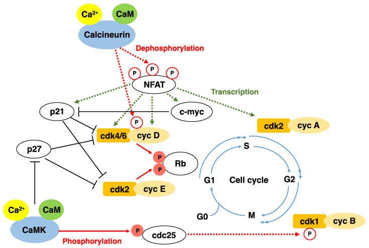
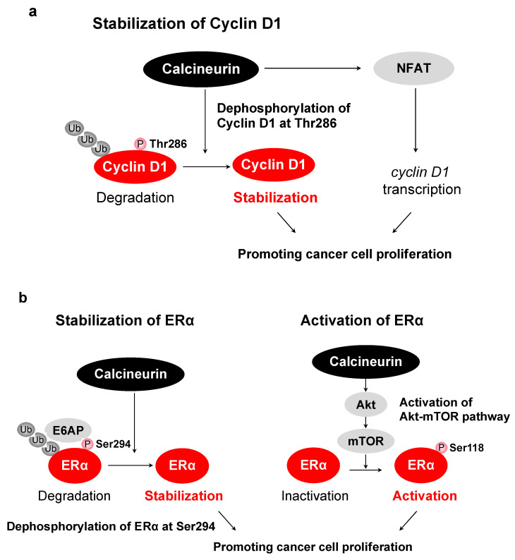
Calcineurin, a calcium-dependent serine/threonine phosphatase, integrates the alterations in intracellular calcium levels into downstream signaling pathways by regulating the phosphorylation states of several targets. Intracellular Ca2+ is essential for normal cellular physiology and cell cycle progression at certain critical stages of the cell cycle. Recently, it was reported that calcineurin is activated in a variety of cancers. Given that abnormalities in calcineurin signaling can lead to malignant growth and cancer, the calcineurin signaling pathway could be a potential target for cancer treatment. For example, NFAT, a typical substrate of calcineurin, activates the genes that promote cell proliferation. Furthermore, cyclin D1 and estrogen receptors are dephosphorylated and stabilized by calcineurin, leading to cell proliferation. In this review, we focus on the cell proliferative functions and regulatory mechanisms of calcineurin and summarize the various substrates of calcineurin. We also describe recent advances regarding dysregulation of the calcineurin activity in cancer cells. We hope that this review will provide new insights into the potential role of calcineurin in cancer development.
Keywords: calcineurin; cancer; cell cycle; dephosphorylation; intracellular calcium ions.
Conflict of interest statement
The authors declare no conflict of interest.
Figures
Similar articles
-
Calcineurin in cancer signaling networks.Nagoya J Med Sci. 2025 May;87(2):182-195. doi: 10.18999/nagjms.87.2.182. Nagoya J Med Sci. 2025. PMID: 40765797 Free PMC article. Review.
-
The Role of Calcium-Calcineurin-NFAT Signaling Pathway in Health and Autoimmune Diseases.Front Immunol. 2020 Mar 10;11:195. doi: 10.3389/fimmu.2020.00195. eCollection 2020. Front Immunol. 2020. PMID: 32210952 Free PMC article. Review.
-
Calcineurin/NFATc1 pathway contributes to cell proliferation in hepatocellular carcinoma.Dig Dis Sci. 2012 Dec;57(12):3184-8. doi: 10.1007/s10620-012-2255-8. Epub 2012 Jun 22. Dig Dis Sci. 2012. PMID: 22722879
-
Dephosphorylation of NFAT by Calcineurin inhibits Skp2-mediated degradation.J Biochem. 2024 Mar 4;175(3):235-244. doi: 10.1093/jb/mvad103. J Biochem. 2024. PMID: 38030387
-
Interaction of calcineurin with substrates and targeting proteins.Trends Cell Biol. 2011 Feb;21(2):91-103. doi: 10.1016/j.tcb.2010.09.011. Epub 2010 Nov 4. Trends Cell Biol. 2011. PMID: 21115349 Free PMC article. Review.
Cited by
-
Calcium signaling mediates proliferation of the precursor cells that give rise to the ciliated left-right organizer in the zebrafish embryo.Front Mol Biosci. 2023 Dec 12;10:1292076. doi: 10.3389/fmolb.2023.1292076. eCollection 2023. Front Mol Biosci. 2023. PMID: 38152112 Free PMC article.
-
RCAN1-mediated calcineurin inhibition as a target for cancer therapy.Mol Med. 2022 Jun 18;28(1):69. doi: 10.1186/s10020-022-00492-7. Mol Med. 2022. PMID: 35717152 Free PMC article. Review.
-
NFAT activation by FKBP52 promotes cancer cell proliferation by suppressing p53.Life Sci Alliance. 2024 May 21;7(8):e202302426. doi: 10.26508/lsa.202302426. Print 2024 Aug. Life Sci Alliance. 2024. PMID: 38803221 Free PMC article.
-
Calcineurin in cancer signaling networks.Nagoya J Med Sci. 2025 May;87(2):182-195. doi: 10.18999/nagjms.87.2.182. Nagoya J Med Sci. 2025. PMID: 40765797 Free PMC article. Review.
-
Ca2+-PP2B-PSD-95 axis: A novel regulatory mechanism of the phosphorylation state of Serine 295 of PSD-95.PLoS One. 2024 Nov 7;19(11):e0313441. doi: 10.1371/journal.pone.0313441. eCollection 2024. PLoS One. 2024. PMID: 39509447 Free PMC article.
References
Publication types
MeSH terms
Substances
Grants and funding
LinkOut - more resources
Full Text Sources
Medical
Research Materials
Miscellaneous
