. 2022 Jan 21;23(3):1163.
doi: 10.3390/ijms23031163.

P-Glycoprotein and Androgen Receptor Expression Reveals Independence of Canine Prostate Cancer from Androgen Hormone Stimulation

Affiliations

P-Glycoprotein and Androgen Receptor Expression Reveals Independence of Canine Prostate Cancer from Androgen Hormone Stimulation

Alexandre Matheus Baesso Cavalca et al. Int J Mol Sci. .

Abstract

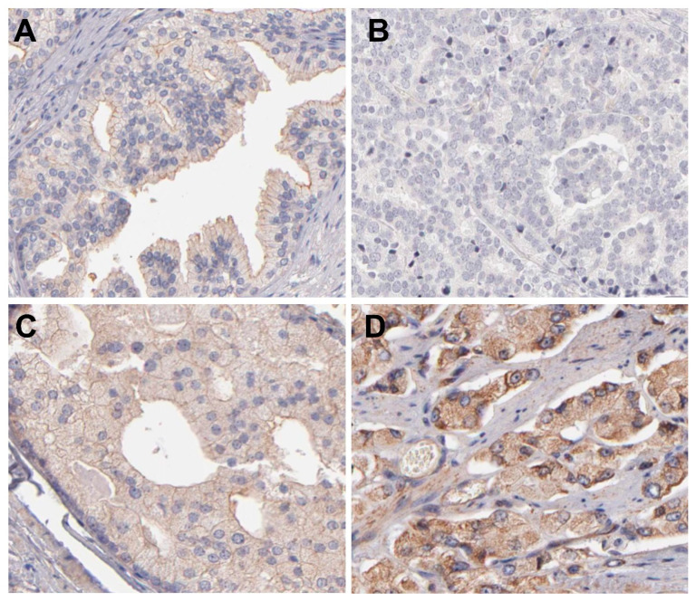
Canine prostate cancer (PC) is an aggressive disease, and dogs can be considered comparative models for human PC. In recent years, canine PC has been shown to resemble human castrate-resistant prostate cancer. The influx and efflux of testosterone in prostatic luminal cells are regulated by P-glycoprotein (P-gp). Therefore, human PC generally lacks P-gp expression and maintains the expression of androgen receptors (ARs). However, this co-expression has not previously been investigated in dogs. Therefore, this study aimed to evaluate AR and P-gp co-expression to elucidate these protein patterns in canine prostate samples. We identified AR/P-gp double immunofluorescence co-expression of both proteins in normal luminal cells. However, in canine PC, cells lack AR expression and exhibit increased P-gp expression. These results were confirmed by gene expression analyses. Overall, our results strongly suggest that normal canine prostate testosterone influx may be regulated by P-gp expression, and that during progression to PC, prostatic cells lack AR expression and P-gp overexpress. P-gp expression in canine PC may be related to a phenotype of multiple drug resistance.

Keywords: ABCB1; comparative oncology; prostatic disease; testosterone.


Conflict of interest statement

The authors declare no conflict of interest.

Figures

Figure 1
Figure 1
Expression of androgen receptor (AR), P-glycoprotein (P-gp) and doubled stained cells (AR/P-gp) in canine prostatic samples. (A): Analysis of variance (ANOVA) revealing high doubled stained cells (AR/P-gp) in normal samples and decreased expression on proliferative inflammatory atrophy (PIA) and prostate cancer (PC) samples. (B): High AR expression in normal samples compared to PIA and PC. (C): ANOVA analysis revealing higher P-gp expression on PC samples, compared to normal and PIA. (D): Positive correlation between the number of AR and P-gp positive stained cells in normal samples, indicating a dependency of both variables. Therefore, in normal prostate, increased AR expression also increases P-gp expression. (E): Correlation analysis revealing that samples with higher AR expression also demonstrate higher P-gp expression in PIA samples. Similar to normal samples, in PIA samples, AR and P-gp expression presents dependency, indicating control of P-gp to androgen hormones influx. (F): No correlation is found between AR and P-gp expression in PC samples. Thus, in canine PC, no dependency or relation is detected between AR and P-gp expression.
Figure 2
Figure 2
Immunofluorescence analysis of androgen receptor (AR) (green color) and P-glycoprotein (P-gp) (red color) in canine prostatic samples. (AD): expression of P-gp (B) and AR (C) in normal prostate samples and merged image (D) demonstrating the doubled stained cells with AR expression in nucleus and P-gp in cell membrane. (EH): expression of P-gp (F), AR (G) and merged image (AR/P-gp) in PIA samples with surrounding normal tissue. The normal prostatic cells are showing positive P-gp expression in the membrane and nuclear AR expression (arrows). Conversely, PIA areas are showing no membranous P-gp and lack of AR expression (nucleus stained with DAPI—blue color) (arrowhead). (IL): Canine PC with solid pattern showing cytoplasmic and membranous P-gp expression (J) and AR cytoplasmic expression (K). In the merged image (AR/P-gp double-stained) (L), it is observed lack of nuclear AR (cells stained with DAPI) and cell cytoplasm with yellowish stained (indicating a colocalization of AR and P-gp in cytoplasm). (MO) and ‘(P): PC samples showing low P-gp expression and high AR cytoplasmic expression with no nuclear expression (PT): PC samples showing P-gp membranous expression with AR stained in nucleus and cytoplasm (TX), and (Z): PC samples revealing membranous P-gp expression with AR cytoplasmic expression (Z). Immunofluorescence images, DAPI counterstaining.
Figure 3
Figure 3
MDR1 and AR gene expression analysis in canine prostatic samples. (A): MDR1 expression revealing higher transcript levels on canine prostate cancer (PC), compared to normal and proliferative inflammatory atrophy (PIA) samples. (B): AR transcripts in canine prostatic samples. A higher AR expression on normal samples, compared to normal and PIA samples is observed. (C): Linear regression analysis revealing a positive association between MDR1 and AR transcripts. (D): Canine PC lacking association between MDR1 and AR transcripts. (E): Linear regression revealing positive association between MDR1 and AR transcripts.
Figure 4
Figure 4
AR immunohistochemical expression human prostatic samples. (A): Normal prostate gland demonstrating strong nuclear AR expression in luminal cells. (B): A high-grade prostate cancer (PC) showing strong nuclear AR expression. (C): A low grade PC sample showing moderate AR nuclear expression. (D): A high-grade PC showing moderate nuclear AR expression. Image credit of the IHC images: Human Protein Atlas, www.proteinatlas.org. Image available at the following URL: https://www.proteinatlas.org/ENSG00000169083-AR/pathology/prostate+cancer, accessed on 16 November 2021.
Figure 5
Figure 5
P-glycoprotein (P-gp) immunohistochemical expression human prostatic samples. (A): Normal prostate gland showing weak membranous P-gp expression by luminal cells. (B): A high-grade prostate cancer (PC) showing no P-gp expression. (C): A PC representing moderate cytoplasmic and membranous P-gp expression. (D): A high-grade PC showing strong membranous and cytoplasmic P-gp expression. Image credit of the IHC images: Human Protein Atlas, www.proteinatlas.org. Image available at the following URL: https://www.proteinatlas.org/ENSG00000085563-ABCB1/pathology/prostate+cancer#img, accessed on 16 November 2021.
Figure 6
Figure 6
MDR1 and AR transcriptional evaluation on human prostate samples. (A): Higher MDR1 gene expression in normal prostatic samples compared to prostate cancer (PC) samples. The normal samples demonstrate higher transcripts per million (TPM) (green dots) than PC (red dots). (B): AR gene expression in normal prostate and PC cases. Higher AR expression on PC samples is observed. The normal samples demonstrate lower TPM (green dots) than PC (red dots). (C): Correlation analysis between MDR1 and AR gene expression in normal prostatic samples showing no association. (D): Positive low correlation between MDR1 and AR transcripts on human PC samples. The gene expression dotplot is generated using the GEPIA database (http://gepia.cancer-pku.cn/about.html, accessed on 16 November 2021). PRAD: PC dataset.

Similar articles

Cited by

References

    1. Griffin J.E. Androgen resistance—The clinical and molecular spectrum. N. Engl. J. Med. 1992;326:611–618. - PubMed
    1. Banerjee P.P., Banerjee S., Brown T.R., Zirkin B.R. Androgen action in prostate function and disease. Am. J. Clin. Exp. Urol. 2018;6:62–77. - PMC - PubMed
    1. Evans R.M. The steroid and thyroid hormone receptor superfamily. Science. 1988;240:889–895. doi: 10.1126/science.3283939. - DOI - PMC - PubMed
    1. Rosner W., Hryb D.J., Khan M.S., Nakhla A.M., Romas N.A. Sex hormone-binding globulin mediates steroid hormone signal transduction at the plasma membrane. J. Steroid Biochem. Mol. Biol. 1999;69:481–485. doi: 10.1016/S0960-0760(99)00070-9. - DOI - PubMed
    1. Giorgi E.P., Stein W.D. The transport of steroids into animal cells in culture. Endocrinology. 1981;108:688–697. doi: 10.1210/endo-108-2-688. - DOI - PubMed

MeSH terms

Substances