. 2022 Jan 21;23(3):1177.
doi: 10.3390/ijms23031177.

Induced Neurodifferentiation of hBM-MSCs through Activation of the ERK/CREB Pathway via Pulsed Electromagnetic Fields and Physical Stimulation Promotes Neurogenesis in Cerebral Ischemic Models

Affiliations

Induced Neurodifferentiation of hBM-MSCs through Activation of the ERK/CREB Pathway via Pulsed Electromagnetic Fields and Physical Stimulation Promotes Neurogenesis in Cerebral Ischemic Models

Hee-Jung Park et al. Int J Mol Sci. .

Abstract

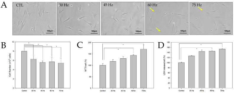
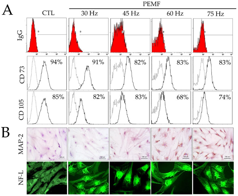
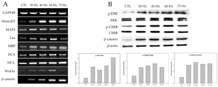
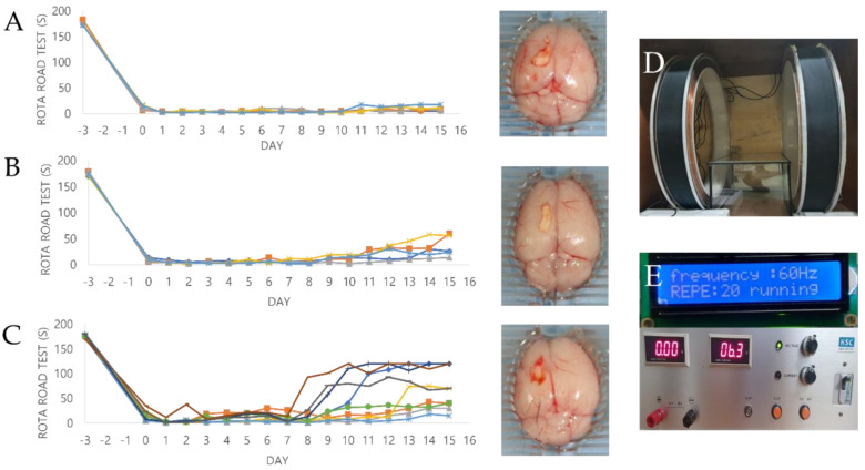
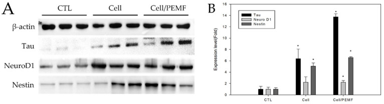
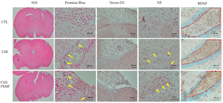
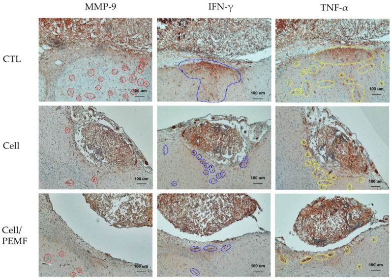
Stroke is among the leading causes of death worldwide, and stroke patients are more likely to live with permanent disabilities even after treatment. Several treatments are being developed to improve the quality of life of patients; however, these treatments still have important limitations. Our study thus sought to evaluate the neural differentiation of human bone marrow mesenchymal stem cells (hBM-MSCs) at various pulsed electromagnetic field (PEMF) frequencies. Furthermore, the effects of selected frequencies in vivo were also evaluated using a mouse ischemia stroke model. Cell proliferation decreased by 20% in the PEMF group, as demonstrated by the 3-[4,5-dimethylthiazol-2-yl]-2,5-diphenyl tetrazolium bromide (MTT) assay, and lactate dehydrogenase (LDH) secretion increased by approximately 10% in an LDH release assay. Fluorescence-activated cell sorting (FACS) analysis demonstrated that CD73 and CD105 were downregulated in the PEMF group at 60 Hz. Moreover, microtubule-associated protein 2 (MAP-2) and neurofilament light chain (NF-L) were upregulated in cell cultures at 60 and 75 Hz. To assess the effects of PEMF in vivo, cerebral ischemia mice were exposed to a PEMF at 60 Hz. Neural-related proteins were significantly upregulated in the PEMF groups compared with the control and cell group. Upon conducting rotarod tests, the cell/PEMF group exhibited significant differences in motor coordination at 13 days post-treatment when compared with the control and stem-cell-treated group. Furthermore, the cell and cell/PEMF group exhibited a significant reduction in the expression of matrix metalloproteinase-9 (MMP-9), tumor necrosis factor-alpha (TNF-α) and interferon-gamma (IFN-γ) in the induced ischemic area compared with the control. Collectively, our findings demonstrated that PEMFs at 60 and 75 Hz could stimulate hBM-MSCs neural differentiation in vitro, in addition to promoting neurogenesis to enhance the functional recovery process by reducing the post-stroke inflammatory reaction.

Keywords: cerebral ischemia stroke; inflammatory cytokines; neural differentiation; pulse electromagnetic fields.


Conflict of interest statement

The authors declare no conflict of interest.

Figures

Figure 1
Figure 1
(A) Morphological changes of hBM-MSCs exposed to PEMF for 3 days (bar = 100 μm). (B) Cell counting was performed using a Scepter cell counter (PHCC00000, Millipore) (n = 3); * p < 0.05: statistically significant difference compared with the control. (C,D) MTT and LDH assays were performed to assess the metabolic activity of hBM-MSCs after three days of PEMF treatment at various frequencies (n = 3); * p < 0.05: statistically significant difference compared with the control.
Figure 2
Figure 2
(A) Results of fluorescence-activated cell sorting (FACS) analysis on hBM-MSCs surface markers (CD73 and CD105) and IgG control after PEMF for three days. (B) Immunohistochemical analysis of MAP-2 antibodies on hBM-MSCs cultured after PEMF for three days (original magnification: 100×); and immunofluorescence staining of NF-L antibodies on hBM-MSCs after PEMF for three days (original magnification: 400×).
Figure 3
Figure 3
(A) Levels of neural gene expression detected by RT-PCR in hBM-MSCs after three days of PEMF treatment. Total cellular mRNA was extracted to conduct RT-PCR. (B) Western blot analysis of hBM-MSCs after PEMF for three days. Total cells lysates were immunoblotted with ERK-, p-ERK-, CREB-, and p-CREB-specific antibodies. β-actin served as an internal control.
Figure 4
Figure 4
This is the result of the Rota-rod test behavioral experiment for 15 days after the ischemia model. All mice were trained daily for 3 days before the stroke modeling for the Rota-rod (3 cm in diameter, 15 rpm) test. To evaluate animal behavior, the Rota-rod test was performed every day for 15 days after transplantation. −3 days = internal baseline before the surgery, 0 days = right after the surgery. (A) Control group (n = 5), (B) Cell group (n = 5), (C) Cell/PEMF group (n = 9). (D) Two solenoid coils. (E) Signal generator.
Figure 5
Figure 5
Western blot analysis after transplantation of hBM-MSCs in the cerebral ischemia mouse model at 15 days. (A) The cell and cell/PEMF group exhibited higher levels of neural protein expression compared to the control group. (B) Quantitative analysis of expression level in comparison to β-actin, an internal control. The Western blot band intensities were measured with the ImageJ software. Each bar represents the mean ± SD of independent experiments performed in triplicate (n = 3). * p < 0.05: significant difference compared with the control group (n = 3).
Figure 6
Figure 6
Cerebral infarction area and immunohistochemical staining 15 days after surgery. Expression of neural-related proteins in the control group, cell group, and cell/PEMF group. Yellow arrow: Prussian Blue and NF expression. Hematoxylin and eosin (H&E) staining (original magnification: 1.15×), Prussian Blue (Original magnification: 200×), Neuro D1 (original magnification: 100×), NF (original magnification: 400×), and BDNF (original magnification: 200×).
Figure 7
Figure 7
Immunohistochemical staining 15 days after surgery. Expression of MMP-9, IFN-γ, and TNF-α in the control, cell, and cell/PEMF groups. Red dotted line: MMP-9 expression; blue dotted line: IFN-γ expression; yellow dotted line: TNF-α expression (original magnification: 100×).

Similar articles

Cited by

References

    1. Avan A., Digaleh H., Di Napoli M., Stranges S., Behrouz R., Shojaeianbabaei G., Amiri A., Tabrizi R., Mokhber N., Spence J.D., et al. Socioeconomic status and stroke incidence, prevalence, mortality, and worldwide burden: An ecological analysis from the Global Burden of Disease Study 2017. BMC Med. 2019;17:191. doi: 10.1186/s12916-019-1397-3. - DOI - PMC - PubMed
    1. Donnan G.A., Fisher M., Macleod M., Davis S.M. Stroke. Lancet. 2008;371:1612–1623. doi: 10.1016/S0140-6736(08)60694-7. - DOI - PubMed
    1. Virani S.S., Alonso A., Benjamin E.J., Bittencourt M.S., Callaway C.W., Carson A.P., Chamberlain A.M., Chang A.R., Cheng S., Delling F.N., et al. Heart Disease and Stroke Statistics-2020 Update: A Report From the American Heart Association. Circulation. 2020;141:e139–e596. doi: 10.1161/CIR.0000000000000757. - DOI - PubMed
    1. Zhang J., Chopp M. Cell-based therapy for ischemic stroke. Expert Opin. Biol. Ther. 2013;13:1229–1240. doi: 10.1517/14712598.2013.804507. - DOI - PMC - PubMed
    1. Salem H.K., Thiemermann C. Mesenchymal stromal cells: Current understanding and clinical status. Stem Cells. 2010;28:585–596. doi: 10.1002/stem.269. - DOI - PMC - PubMed

MeSH terms

Substances