

TRIM37 Promotes Pancreatic Cancer Progression through Modulation of Cell Growth, Migration, Invasion, and Tumor Immune Microenvironment
- PMID: 35163097
- PMCID: PMC8835669
- DOI: 10.3390/ijms23031176
TRIM37 Promotes Pancreatic Cancer Progression through Modulation of Cell Growth, Migration, Invasion, and Tumor Immune Microenvironment
Abstract
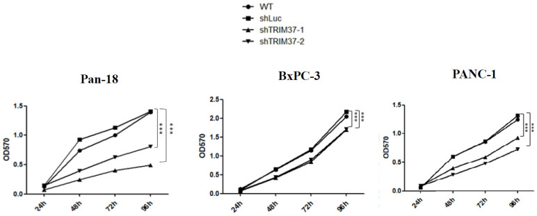
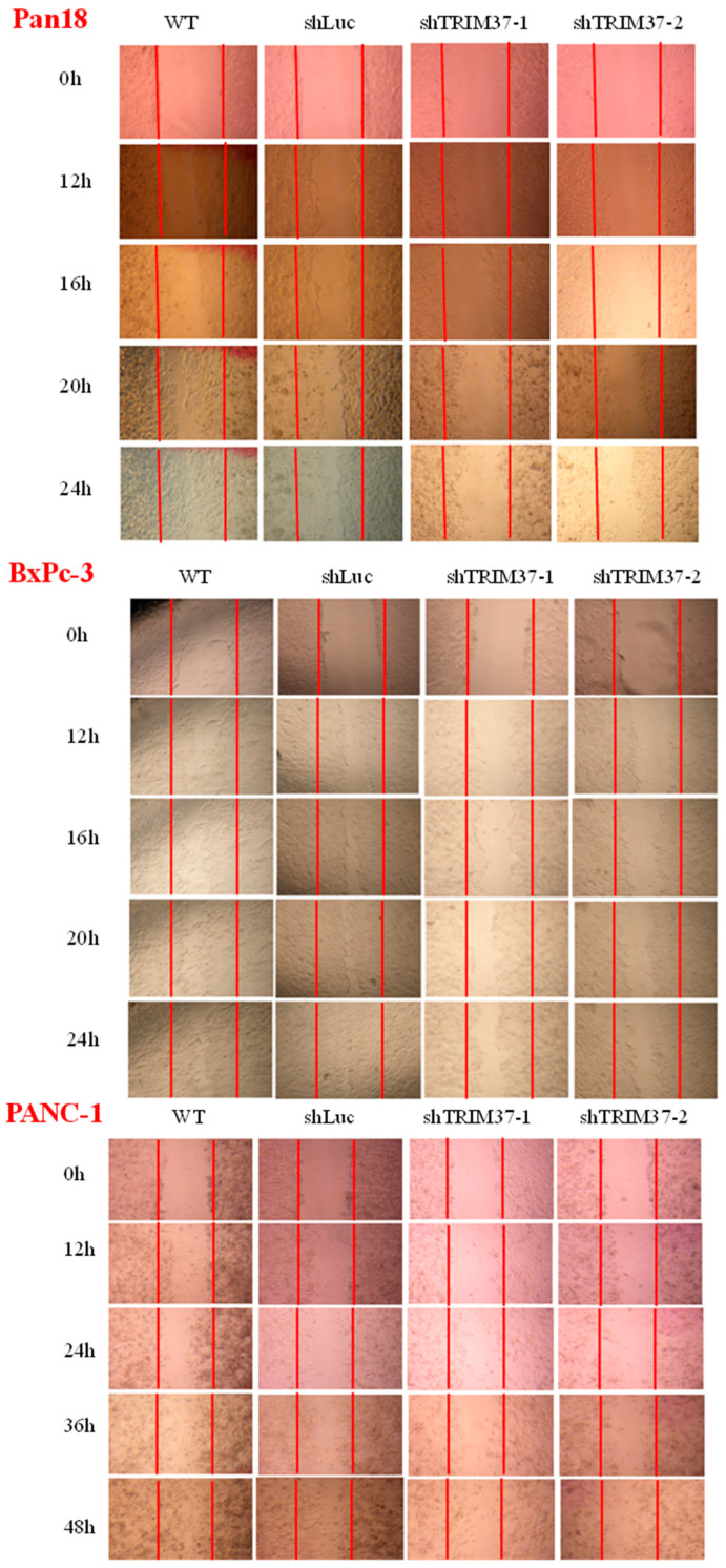
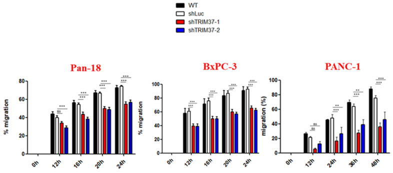
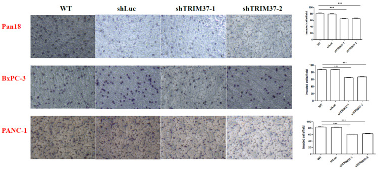
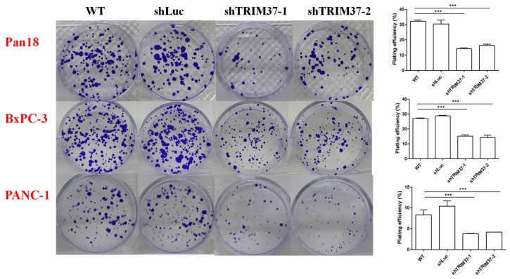
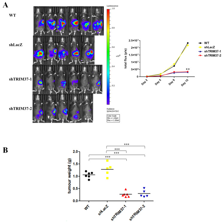
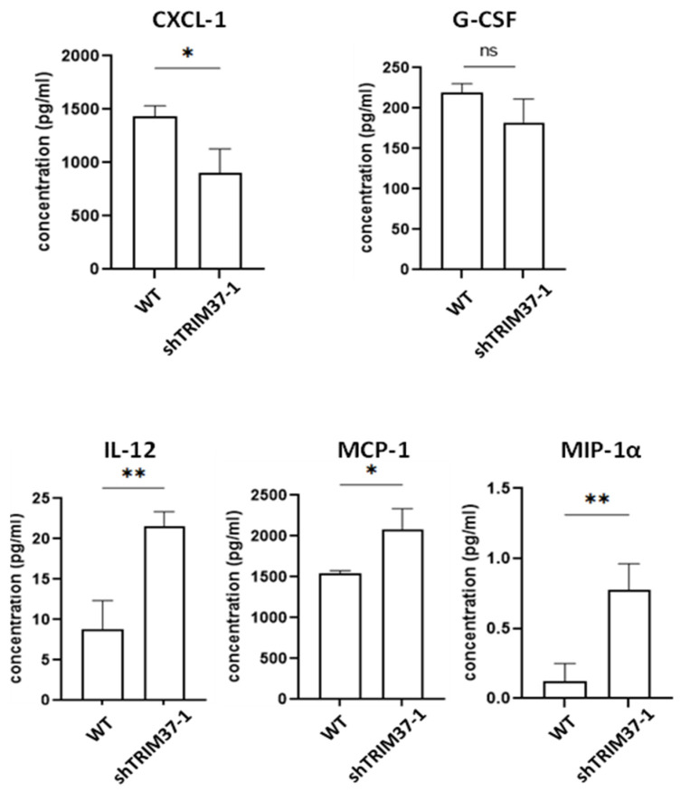
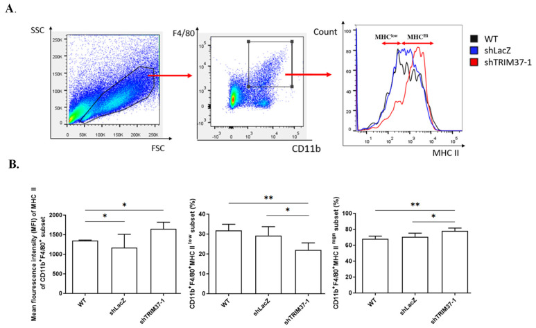
TRIM37 dysregulation has been observed in several cancer types, implicating its possible role in tumorigenesis. However, the role of TRIM37 in pancreatic cancer progression remains unclear. In the present study, we observed that TRIM37 knockdown resulted in reduced proliferation, clonogenicity, migration, and invasion ability of pancreatic cancer cells. Furthermore, an in vivo study using an orthotopic syngeneic animal model further confirmed that reduced expression of TRIM37 in cancer cells suppressed tumor growth in vivo. Moreover, in mice bearing TRIM37 knockdown pancreatic cancer cells, the proportion of CD11b+F4/80+MHCIIlow immunosuppressive macrophages was significantly reduced in tumor milieu, which might be due to the regulatory role of TRIM37 in cytokine production by pancreatic cancer cells. Collectively, these findings suggest a key role of TRIM37 in promoting pancreatic cancer progression.
Keywords: TRIM37; immune microenvironment; pancreatic cancer.
Conflict of interest statement
The authors declare no conflict of interest.
Figures
Similar articles
-
TRIM37 interacts with EZH2 to epigenetically suppress PTCH1 and regulate stemness in glioma stem cells through sonic hedgehog pathway.J Neurooncol. 2024 Sep;169(2):269-279. doi: 10.1007/s11060-024-04726-y. Epub 2024 Jun 17. J Neurooncol. 2024. PMID: 38884661
-
lncRNA NUTM2A-AS1 Targets the SRSF1/Trim37 Signaling Pathway to Promote the Proliferation and Invasion of Breast Cancer.Comput Math Methods Med. 2022 Aug 2;2022:3299336. doi: 10.1155/2022/3299336. eCollection 2022. Comput Math Methods Med. 2022. Retraction in: Comput Math Methods Med. 2023 Sep 27;2023:9895079. doi: 10.1155/2023/9895079. PMID: 35959349 Free PMC article. Retracted.
-
TRIM37 promotes the aggressiveness of ovarian cancer cells and increases c-Myc expression by binding to HUWE1.Arch Biochem Biophys. 2022 Oct 15;728:109372. doi: 10.1016/j.abb.2022.109372. Epub 2022 Jul 31. Arch Biochem Biophys. 2022. PMID: 35921902
-
TRIM37: a critical orchestrator of centrosome function.Cell Cycle. 2021 Dec;20(23):2443-2451. doi: 10.1080/15384101.2021.1988289. Epub 2021 Oct 21. Cell Cycle. 2021. PMID: 34672905 Free PMC article. Review.
-
Epigenetic regulation of the tumor microenvironment: A leading force driving pancreatic cancer.Pancreatology. 2024 Sep;24(6):878-886. doi: 10.1016/j.pan.2024.07.005. Epub 2024 Jul 15. Pancreatology. 2024. PMID: 39095296 Review.
Cited by
-
Comprehensive Characterization of a Novel E3-Related Gene Signature With Implications in Prognosis and Immunotherapy of Low-Grade Gliomas.Front Genet. 2022 Jun 27;13:905047. doi: 10.3389/fgene.2022.905047. eCollection 2022. Front Genet. 2022. PMID: 35832194 Free PMC article.
-
KIFC1 depends on TRIM37-mediated ubiquitination of PLK4 to promote centrosome amplification in endometrial cancer.Cell Death Discov. 2024 Sep 30;10(1):419. doi: 10.1038/s41420-024-02190-1. Cell Death Discov. 2024. PMID: 39349439 Free PMC article.
-
Exploiting E3 ubiquitin ligases to reeducate the tumor microenvironment for cancer therapy.Exp Hematol Oncol. 2023 Mar 30;12(1):34. doi: 10.1186/s40164-023-00394-2. Exp Hematol Oncol. 2023. PMID: 36998063 Free PMC article. Review.
-
N6-methyladenosine-modified TRIM37 augments sunitinib resistance by promoting the ubiquitin-degradation of SmARCC2 and activating the Wnt signaling pathway in renal cell carcinoma.Cell Death Discov. 2024 Sep 30;10(1):418. doi: 10.1038/s41420-024-02187-w. Cell Death Discov. 2024. PMID: 39349442 Free PMC article.
References
-
- Brembeck F.H., Schreiber F.S., Deramaudt T.B., Craig L., Rhoades B., Swain G., Grippo P., Stoffers A.D., Silberg D.G., Rustgi A.K. The mutant K-ras oncogene causes pancreatic periductal lymphocytic infiltration and gastric mucous neck cell hyperplasia in transgenic mice. Cancer Res. 2003;63:2005–2009. - PubMed
MeSH terms
Substances
Grants and funding
LinkOut - more resources
Full Text Sources
Medical
Research Materials
