Review
. 2022 Jan 22;23(3):1232.
doi: 10.3390/ijms23031232.

Prion Protein: The Molecule of Many Forms and Faces

Affiliations
Review

Prion Protein: The Molecule of Many Forms and Faces

Valerija Kovač et al. Int J Mol Sci. .

Abstract

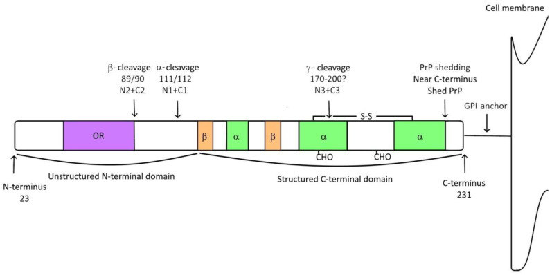
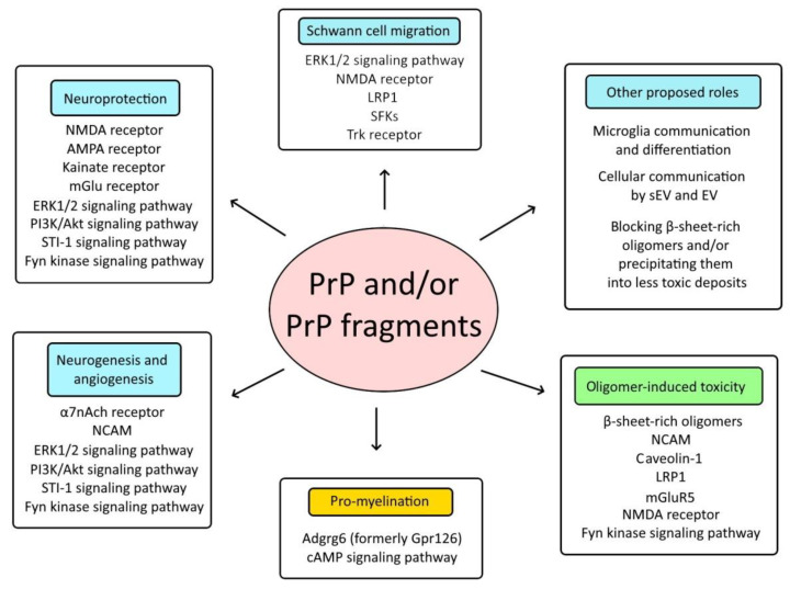
Cellular prion protein (PrPC) is a glycosylphosphatidylinositol (GPI)-anchored protein most abundantly found in the outer membrane of neurons. Due to structural characteristics (a flexible tail and structured core), PrPC interacts with a wide range of partners. Although PrPC has been proposed to be involved in many physiological functions, only peripheral nerve myelination homeostasis has been confirmed as a bona fide function thus far. PrPC misfolding causes prion diseases and PrPC has been shown to mediate β-rich oligomer-induced neurotoxicity in Alzheimer's and Parkinson's disease as well as neuroprotection in ischemia. Upon proteolytic cleavage, PrPC is transformed into released and attached forms of PrP that can, depending on the contained structural characteristics of PrPC, display protective or toxic properties. In this review, we will outline prion protein and prion protein fragment properties as well as overview their involvement with interacting partners and signal pathways in myelination, neuroprotection and neurodegenerative diseases.

Keywords: ischemic stroke; myelination; neurodegenerative disease; neuroprotection; prion protein; prion protein fragments.


Conflict of interest statement

The authors declare no conflict of interest.

Figures

Figure 1
Figure 1
Schematic presentation of PrPC with associated cleavages. Mature PrPC is approximately 210 amino acids long. The flexible unstructured N-terminal part (residues 23–120) contains the octapeptide repeat region (OR, purple) whereas the highly structured C-terminal part (residues 121–231) is composed of three α-helices (green), two β-sheets (orange), a disulfide bond, two N-glycans (CHO; positions 181 and 197) and a C-terminal GPI anchor. PrP can undergo four cleavages: α-cleavage (cleavage site position 111/112); β-cleavage (cleavage site position 89/90); γ-cleavage (cleavage site presumably between positions 170–120); and shedding (near the C-terminus of PrP). Cleavages result in released (N1, N2, N3, shed PrP) and attached (C1, C2, C3) fragments of PrPC.
Figure 2
Figure 2
Proteins, signaling pathways and interactions that may be affected by PrP and/or PrP fragments. This scheme presents various proteins, signaling pathways and interactions that reportedly involve PrP and/or its fragments. In ischemic stroke, PrP species were found to be involved in modulating neuroprotection, neurite outgrowth, neurogenesis and angiogenesis. In neurodegenerative diseases, released PrP fragments may act protectively whereas anchored PrP regulates oligomer-induced toxicity. PrP and its derivatives are also involved in Adgrg6-induced myelination homeostasis (orange) and may be involved in microglia communication and differentiation as well as regulating intercellular communication through EVs and sEVs, etc. Several of the proposed interplays are regulated by a direct interaction with PrP species whereas others are regulated indirectly. Protective pathways and interactions are colored blue whereas green color presents harmful outcomes.

References

    1. Scheckel C., Aguzzi A. Prions, prionoids and protein misfolding disorders. Nat. Rev. Genet. 2018;19:405–418. doi: 10.1038/s41576-018-0011-4. - DOI - PubMed
    1. O’Carroll A., Coyle J., Gambin Y. Prions and Prion-like assemblies in neurodegeneration and immunity: The emergence of universal mechanisms across health and disease. Semin. Cell Dev. Biol. 2020;99:115–130. doi: 10.1016/j.semcdb.2019.11.012. - DOI - PubMed
    1. Ritchie D.L., Barria M.A. Prion Diseases: A Unique Transmissible Agent or a Model for Neurodegenerative Diseases? Biomolecules. 2021;11:207. doi: 10.3390/biom11020207. - DOI - PMC - PubMed
    1. Herms J., Tings T., Gall S., Madlung A., Giese A., Siebert H., Schurmann P., Windl O., Brose N., Kretzschmar H. Evidence of presynaptic location and function of the prion protein. J. Neurosci. 1999;19:8866–8875. doi: 10.1523/JNEUROSCI.19-20-08866.1999. - DOI - PMC - PubMed
    1. Bendheim P.E., Brown H.R., Rudelli R.D., Scala L.J., Goller N.L., Wen G.Y. Nearly ubiquitous tissue distribution of the scrapie agent precursor protein. Neurology. 1992;42:149. doi: 10.1212/WNL.42.1.149. - DOI - PubMed