Review
. 2022 Jan 23;23(3):1244.
doi: 10.3390/ijms23031244.

The Role of Biomaterials in Peripheral Nerve and Spinal Cord Injury: A Review

Affiliations
Review

The Role of Biomaterials in Peripheral Nerve and Spinal Cord Injury: A Review

Ben Kaplan et al. Int J Mol Sci. .

Abstract

Peripheral nerve and spinal cord injuries are potentially devastating traumatic conditions with major consequences for patients' lives. Severe cases of these conditions are currently incurable. In both the peripheral nerves and the spinal cord, disruption and degeneration of axons is the main cause of neurological deficits. Biomaterials offer experimental solutions to improve these conditions. They can be engineered as scaffolds that mimic the nerve tissue extracellular matrix and, upon implantation, encourage axonal regeneration. Furthermore, biomaterial scaffolds can be designed to deliver therapeutic agents to the lesion site. This article presents the principles and recent advances in the use of biomaterials for axonal regeneration and nervous system repair.

Keywords: biomaterials; peripheral nerve injury; spinal cord injury.


Conflict of interest statement

The authors declare no conflict of interest. The funders had no role in the design of the study; in the collection, analyses, or interpretation of data; in the writing of the manuscript, or in the decision to publish the results.

Figures

Figure 1
Figure 1
Inner structures of a peripheral nerve. The entire nerve is surrounded by the epineurium collagenous membrane. Each fascicle of axons is separated from surrounding tissue by its own perineurium sheath. Each single axon, along with its myelin sheath, is coated by a delicate endoneurium membrane. Created with BioRender.com.
Figure 2
Figure 2
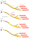
Axonal degeneration and regeneration in peripheral nerve injuries. (A) Cell bodies of spinal motor neurons reside in the spinal cord and extend axons throughout intact peripheral nerves to innervate skeletal muscle fibers. (B) Following transection of axons, axonal segments located distal to the lesion site will undergo Wallerian degeneration. (C) Following axonal degeneration, spinal motor neurons and skeletal muscle fibers may survive; however, the connectivity between these groups of cells is lost. (D) Depending on the severity of injury, regeneration and partial or complete reinnervation of skeletal muscle fibers may take place. Created with BioRender.com.
Figure 3
Figure 3
Axonal degeneration following spinal cord injury. (A) In the intact spinal cord, supra spinal neurons, located in the brain, extend long tract axons into the spinal cord. These axons synapse onto spinal neurons, which extend axons into the peripheral nervous system. (B) When the spinal cord sustains an injury, axons are severed and undergo Wallerian degeneration distal to the lesion site. (C) Following degeneration, supra spinal neurons and spinal neurons often survive; however, their connections are lost. In contrast to peripheral axons, injured axons in the spinal cord do not exhibit meaningful regeneration across the lesion site. Created with BioRender.com.
Figure 4
Figure 4
Types of biomaterial implants for nerve injuries. Hydrogels are injectable biomaterials that can solidify upon implantation and conform to irregular lesion sites. Hollow tube conduits are often used in peripheral nerve injuries; however, they do not contain inner structures to mimic the collagenous membrane of nerve tissues. Porous scaffolds provide better mechanical stability and longer degradation periods compared with hydrogels. Their inner architecture can be designed to mimic the nerve tissue ECM. Created with BioRender.com.
Figure 5
Figure 5
Examples of biomaterial scaffolds for spinal cord and peripheral nerve injuries. (A) differences in size of a foreign body encapsulation around an agarose scaffold (left) and a polyethylene glycol (PEG)- Gelatin methacrylate (GelMA) scaffold implanted in a complete spinal cord injury model in rats [91]. Reproduced with permission. Copyright 2019, Springer Nature. (B) Example of polyester microchannel scaffold. The structure was scanned and imaged using micro-CT [62]. Reproduced with permission. Copyright 2020, Elsevier. (C) Example of a fibrous scaffold for spinal cord repair composed from GelMA and fabricated using electrospinning [99]. Reproduced with permission. Copyright 2019, John Wiley and Sons. (D) neural stem cell derived axons which grow inside a single microchannel adopt a linear growth pattern according to the scaffold topography [91]. Reproduced with permission. Copyright 2019, Springer Nature. (E) Bioprinted alginate scaffold containing accurately deposited neural progenitor cells in each microchannel [100]. Reproduced with permission. Copyright 2018, John Wiley and Sons. (F) 3D printed PEG-GelMA scaffold for spinal cord repair inspired by the native architecture of the spinal cord. (G) Upon implantation NF200 positive axons penetrate the 3D printed microchannels [91]. Reproduced with permission. Copyright 2019, Springer Nature. (H) Anatomically inspired 3D printed scaffold to reconstruct the sciatic nerve bifurcation. (I) In each branch, a different growth factor gradient is located to match the specific type of axons for each pathway. (J) In Vivo implantation of the anatomical scaffold [101]. Reproduced with permission. Copyright 2015, John Wiley and Sons.

References

    1. Kim B.S., Baez C.E., Atala A. Biomaterials for tissue engineering. World J. Urol. 2000;18:2–9. doi: 10.1007/s003450050002. - DOI - PubMed
    1. Naderi H., Matin M.M., Bahrami A.R. Review paper: Critical issues in tissue engineering: Biomaterials, cell sources, angiogenesis, and drug delivery systems. J. Biomater. Appl. 2011;26:383–417. doi: 10.1177/0885328211408946. - DOI - PubMed
    1. Carriel V., Alaminos M., Garzón I., Campos A., Cornelissen M. Tissue engineering of the peripheral nervous system. Expert Rev. Neurother. 2014;14:301–318. doi: 10.1586/14737175.2014.887444. - DOI - PubMed
    1. Boni R., Ali A., Shavandi A., Clarkson A.N. Current and novel polymeric biomaterials for neural tissue engineering. J. Biomed. Sci. 2018;25:90. doi: 10.1186/s12929-018-0491-8. - DOI - PMC - PubMed
    1. Park E., Velumian A.A., Fehlings M.G. The Role of Excitotoxicity in Secondary Mechanisms of Spinal Cord Injury: A Review with an Emphasis on the Implications for White Matter Degeneration. J. Neurotrauma. 2004;21:754–774. doi: 10.1089/0897715041269641. - DOI - PubMed

MeSH terms

Substances