. 2022 Jan 24;23(3):1286.
doi: 10.3390/ijms23031286.

Unravelling Mechanisms of Doxorubicin-Induced Toxicity in 3D Human Intestinal Organoids

Affiliations

Unravelling Mechanisms of Doxorubicin-Induced Toxicity in 3D Human Intestinal Organoids

Daniela Rodrigues et al. Int J Mol Sci. .

Abstract

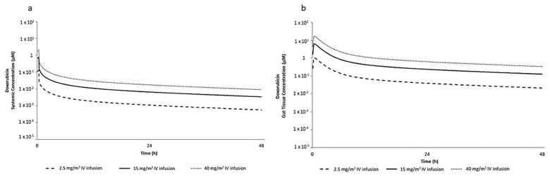
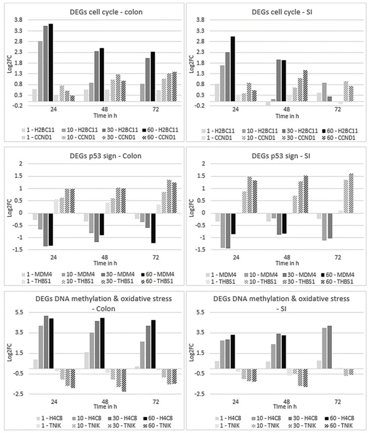
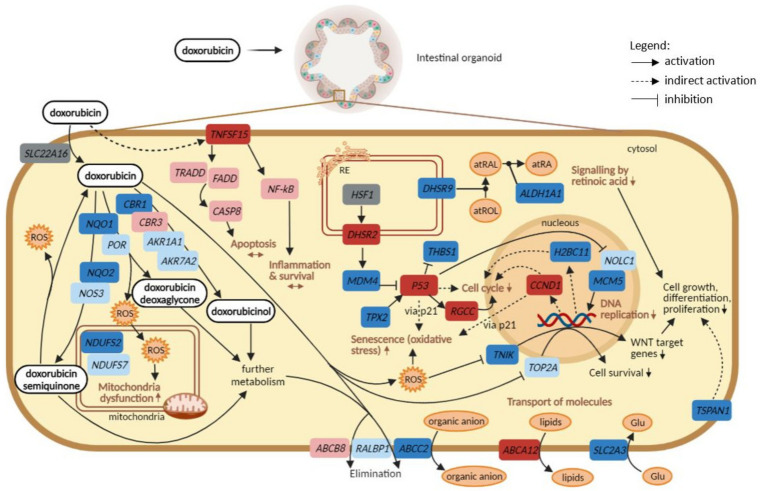
Doxorubicin is widely used in the treatment of different cancers, and its side effects can be severe in many tissues, including the intestines. Symptoms such as diarrhoea and abdominal pain caused by intestinal inflammation lead to the interruption of chemotherapy. Nevertheless, the molecular mechanisms associated with doxorubicin intestinal toxicity have been poorly explored. This study aims to investigate such mechanisms by exposing 3D small intestine and colon organoids to doxorubicin and to evaluate transcriptomic responses in relation to viability and apoptosis as physiological endpoints. The in vitro concentrations and dosing regimens of doxorubicin were selected based on physiologically based pharmacokinetic model simulations of treatment regimens recommended for cancer patients. Cytotoxicity and cell morphology were evaluated as well as gene expression and biological pathways affected by doxorubicin. In both types of organoids, cell cycle, the p53 signalling pathway, and oxidative stress were the most affected pathways. However, significant differences between colon and SI organoids were evident, particularly in essential metabolic pathways. Short time-series expression miner was used to further explore temporal changes in gene profiles, which identified distinct tissue responses. Finally, in silico proteomics revealed important proteins involved in doxorubicin metabolism and cellular processes that were in line with the transcriptomic responses, including cell cycle and senescence, transport of molecules, and mitochondria impairment. This study provides new insight into doxorubicin-induced effects on the gene expression levels in the intestines. Currently, we are exploring the potential use of these data in establishing quantitative systems toxicology models for the prediction of drug-induced gastrointestinal toxicity.

Keywords: doxorubicin; human organoid models; molecular mechanisms; toxicity; transcriptomics.


Conflict of interest statement

The authors declare no conflict of interest. The funders had no role in the design of the study; in the collection, analyses, or interpretation of data; in the writing of the manuscript, or in the decision to publish the results.

Figures

Figure 1
Figure 1
Predicted (a) mean total systemic plasma concentration and (b) mean total gut tissue concentration of DOX following 2.5, 15 and 40 mg/m2 intravenous (IV) dose infused over 20 min in humans.
Figure 2
Figure 2
Functional assessment of healthy colon: (a) viability and (b) caspase 3/7 activation; and of SI organoids: (c) viability and (d) caspase 3/7 activation, when exposed to 1, 10, 30 and 60 µM DOX for 24 h in light grey, 48 h in dark grey and 72 h in black, compared with untreated controls. Values are in % of Luminescence. SD was calculated for each condition. Ctrl, control; DOX, doxorubicin; SD, standard deviation; SI, small intestine; Unt, untreated; Veh, vehicle. * p value of 0.03; ** p value of 0.002; *** p value of 0.0004; **** p value of 0.0001.
Figure 3
Figure 3
Morphological changes assessed through imaging analysis of healthy colon: (a) size, (b) percentage of cell death, and (e) microscope image analysis; and SI organoids: (c) size, (d) percentage of cell death, and (f) microscope image analysis, when exposed to 1, 10, 30 and 60 µM DOX for 24 h in light grey, 48 h in dark grey and 72 h in black, compared with untreated controls. Values are in % based on fluorescent intensity for each measured parameter. SD was calculated for each condition. Ctrl, control; DOX, doxorubicin; SD, standard deviation; SI, small intestine; Unt, untreated; Veh, vehicle. * p value of 0.04; ** p value of 0.008; *** p value of 0.0009; **** p value of 0.0001. Staining in control wells: Phalloidin-FITC (actin, in red) and Hoechst (DAPI channel, nuclei, in blue); treated wells: DOX bound to the nuclei (TRITC channel, nuclei, in blue); 1 pixel is 3.25 µm.
Figure 4
Figure 4
Gene plots representing the expression profiles of genes involved in cell cycle, the p53 signalling pathway, DNA methylation, and oxidative stress-induced senescence, after 24, 48 and 72 h exposure to all DOX concentrations of organoids derived from colon (on the left) and SI (on the right). Values for gene profiles are based on the log2FC. Plot colours correspond to the different concentrations of DOX: lighter grey represents 1 µM; grey represents 10 µM; dark grey represents 30 µM; black represents 60 µM; and DEG: with or without stripes.
Figure 5
Figure 5
Network of direct targets (red, from Table 4) and their first-degree interactors (blue) affected by DOX. The topology of the network implicates a systemic effect of the drug.
Figure 6
Figure 6
Comparison between transcriptomics and proteomics data, starting from DOX entrance to the cell to the several biological pathways and DEGs that are perturbed. For the alterations in the gene expression levels, concentrations of 30 and 60 µM were considered, at every time point. Genes in dark blue, significantly downregulated; light blue, not significantly downregulated; dark red, significantly upregulated; light red, not significantly upregulated; grey, not available. Image created with BioRender.com (accessed on 23 April 2021).

Similar articles

Cited by

References

    1. Arcamone F., Cassinelli G., Fantini G., Grein A., Orezzi P., Pol C., Spalla C. Adriamycin, 14-hydroxydaunomycin, a new antitumor antibiotic from S. peucetius var. caesius. Biotechnol. Bioeng. 1969;11:1101–1110. doi: 10.1002/bit.260110607. - DOI - PubMed
    1. Carvalho C., Santos R.X., Cardoso S., Correia S., Oliveira P.J., Santos M.S., Moreira P.I. Doxorubicin: The good, the bad and the ugly effect. Curr. Med. Chem. 2009;16:3267–3285. doi: 10.2174/092986709788803312. - DOI - PubMed
    1. Pugazhendhi A., Edison T., Velmurugan B.K., Jacob J.A., Karuppusamy I. Toxicity of Doxorubicin (Dox) to different experimental organ systems. Life Sci. 2018;200:26–30. doi: 10.1016/j.lfs.2018.03.023. - DOI - PubMed
    1. Thorn C.F., Oshiro C., Marsh S., Hernandez-Boussard T., McLeod H., Klein T.E., Altman R.B. Doxorubicin pathways: Pharmacodynamics and adverse effects. Pharmacogenet. Genom. 2011;21:440–446. doi: 10.1097/FPC.0b013e32833ffb56. - DOI - PMC - PubMed
    1. Damiani R.M., Moura D.J., Viau C.M., Caceres R.A., Henriques J.A.P., Saffi J. Pathways of cardiac toxicity: Comparison between chemotherapeutic drugs doxorubicin and mitoxantrone. Arch. Toxicol. 2016;90:2063–2076. doi: 10.1007/s00204-016-1759-y. - DOI - PubMed