

Cellular and Molecular Nature of Fragmentation of Human Embryos
- PMID: 35163271
- PMCID: PMC8836137
- DOI: 10.3390/ijms23031349
Cellular and Molecular Nature of Fragmentation of Human Embryos
Abstract
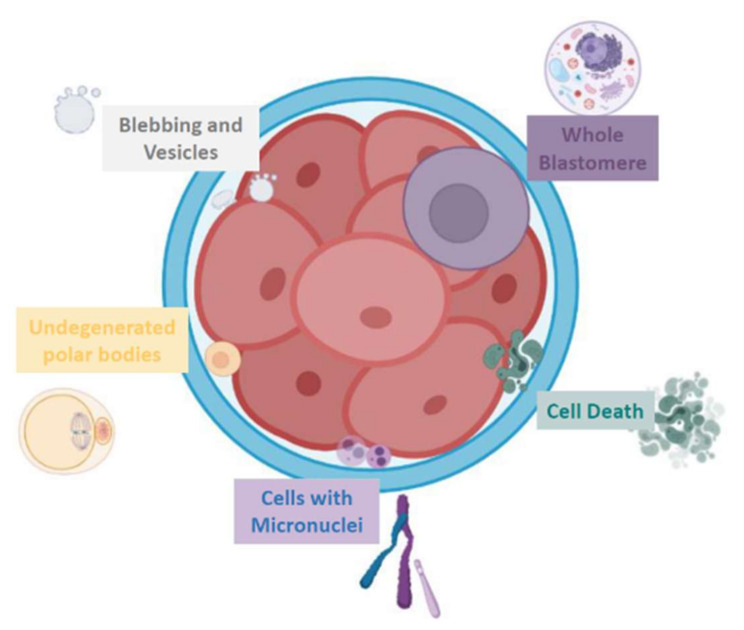
Embryo fragmentation represents a phenomenon generally characterized by the presence of membrane-bound extracellular cytoplasm into the perivitelline space. Recent evidence supports the cellular and molecular heterogeneity of embryo fragments. In this narrative review, we described the different embryo fragment-like cellular structures in their morphology, molecular content, and supposed function and have reported the proposed theories on their origin over the years. We identified articles related to characterization of embryo fragmentation with a specific literature search string. The occurrence of embryo fragmentation has been related to various mechanisms, of which the most studied are apoptotic cell death, membrane compartmentalization of altered DNA, cytoskeletal disorders, and vesicle formation. These phenomena are thought to result in the extrusion of entire blastomeres, release of apoptotic bodies and other vesicles, and micronuclei formation. Different patterns of fragmentation may have different etiologies and effects on embryo competence. Removal of fragments from the embryo before embryo transfer with the aim to improve implantation potential should be reconsidered on the basis of the present observations.
Keywords: apoptosis; embryo; fragmentation; micronuclei; vesicles.
Conflict of interest statement
The authors declare no conflict of interest.
Figures
References
-
- Kim S.-G., Kim Y.-Y., Park J.-Y., Kwak S.-J., Yoo C.-S., Park I.-H., Sun H.-G., Kim J.-W., Lee K.-H., Park H.-D., et al. Early fragment removal on in vitro fertilization day 2 significantly improves the subsequent development and clinical outcomes of fragmented human embryos. Clin. Exp. Reprod. Med. 2018;45:122–128. doi: 10.5653/cerm.2018.45.3.122. - DOI - PMC - PubMed
Publication types
MeSH terms
LinkOut - more resources
Full Text Sources
Other Literature Sources
