Review
. 2022 Jan 25;23(3):1353.
doi: 10.3390/ijms23031353.

Wnt and PI3K/Akt/mTOR Survival Pathways as Therapeutic Targets in Glioblastoma

Affiliations
Review

Wnt and PI3K/Akt/mTOR Survival Pathways as Therapeutic Targets in Glioblastoma

Amir Barzegar Behrooz et al. Int J Mol Sci. .

Abstract

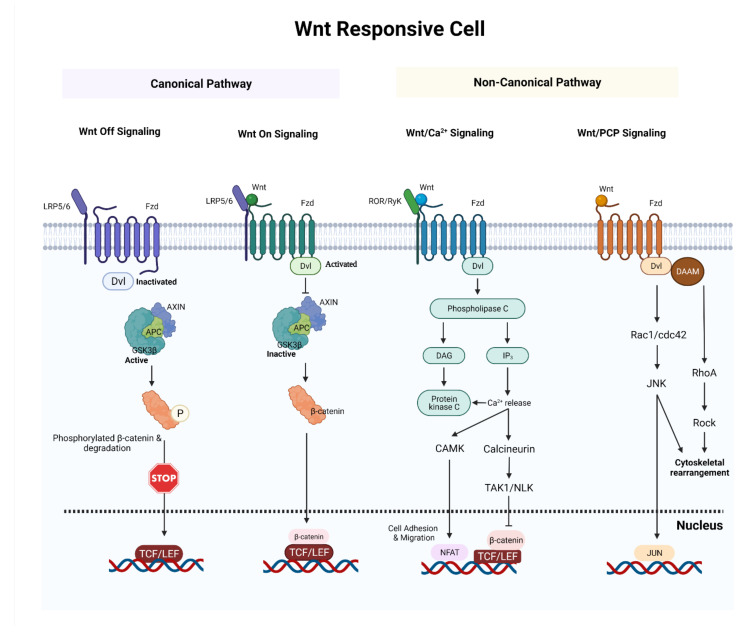
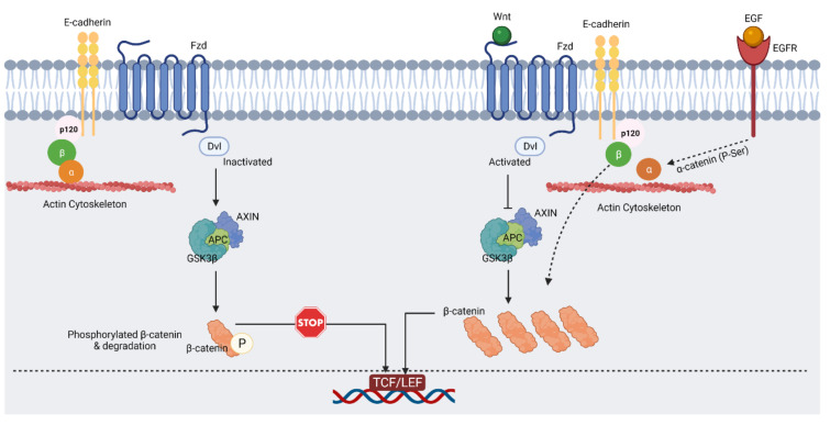
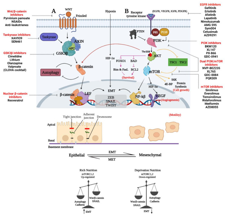
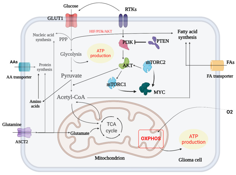
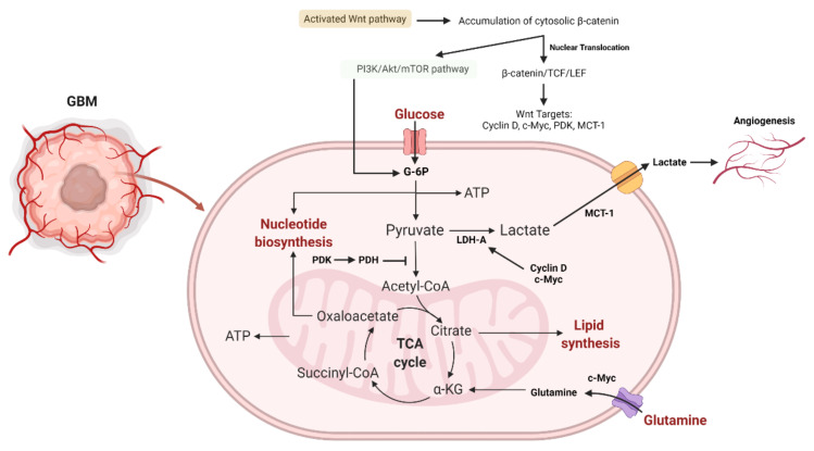
Glioblastoma (GBM) is a devastating type of brain tumor, and current therapeutic treatments, including surgery, chemotherapy, and radiation, are palliative at best. The design of effective and targeted chemotherapeutic strategies for the treatment of GBM require a thorough analysis of specific signaling pathways to identify those serving as drivers of GBM progression and invasion. The Wnt/β-catenin and PI3K/Akt/mTOR (PAM) signaling pathways are key regulators of important biological functions that include cell proliferation, epithelial-mesenchymal transition (EMT), metabolism, and angiogenesis. Targeting specific regulatory components of the Wnt/β-catenin and PAM pathways has the potential to disrupt critical brain tumor cell functions to achieve critical advancements in alternative GBM treatment strategies to enhance the survival rate of GBM patients. In this review, we emphasize the importance of the Wnt/β-catenin and PAM pathways for GBM invasion into brain tissue and explore their potential as therapeutic targets.

Keywords: GBM survival; PI3K/Akt/mTOR; Wnt/β-catenin; autophagy; glioblastoma.


Conflict of interest statement

The authors declare no competing interests.

Figures

Figure 1
Figure 1
A synopsis of the canonical and noncanonical Wnt signaling pathways. In canonical Wnt off signaling, a combination of AXIN and APC allows GSK3β to phosphorylate β-catenin and target it for proteolysis. In canonical Wnt on signaling, Wnt ligands bind and activate Fzd receptor and activated Fzd receptor recruits Dvl protein and AXIN. This blocks the formation of an AXIN–APC complex and inhibits GSK3β. As a result, β-catenin avoids destruction and accumulates in the nucleus. In non-canonical Wnt/Ca2+ signaling, the interaction of Wnt/Fzd with ROR/Ryk co-receptor leads to the formation of IP3 and DAG. DAG activates PKC. IP3 triggers ER Ca2+ release which stimulates calcineurin and CAMKⅡ. This activates TAK1/NLK and NFAT. Activated TAK1/NLK can inhibit TCF/β-catenin signaling and NFAT can regulate cell adhesion and migration. In non-canonical Wnt/PCP signaling, the interaction of Wnt/Fzd leads to the recruitment of Dvl, which utilizes its domains (PDZ and DIX) to produce a complex with DAAM. DAAM then stimulates RhoA. Activated RhoA can activate Rock. Dvl can also produce a complex with Rac1 to activate JNK. Abbreviations: APC, adenomatous polyposis coli; GSK3β, glycogen synthase kinase 3β; LRP5/6, low-density lipoprotein receptor-related protein 5/6; TCF/LEF, T-cell factor/lymphoid enhancer-binding factor; IP3, inositol trisphosphate; DAG, diacylglycerol; CAMKⅡ, calcium/calmodulin-dependent protein kinase II; TAK1, transforming growth factor beta-activated kinase 1; NLK, nemo like kinase; NFAT, nuclear factor of activated T-Cells; Dvl; disheveled; Fzd, frizzled; DAAM, disheveled-associated activator of morphogenesis, ROCK, Rho-associated kinase. (Created with https://biorender.com/, accessed on 18 December 2021).
Figure 2
Figure 2
In epithelial cell–cell adhesion and Wnt signaling, the E-cadherin/β-catenin complex plays a critical role. In the absence of Wnt ligand (on the left), activated GSK3β can phosphorylate β-catenin, which leads to its proteasomal degradation. As result of that, β-catenin remains in the cadherin/β-catenin/α-catenin complex that is attached to the cytoskeleton (created with https://biorender.com/, accessed on 12 September 2021). In the presence of Wnt ligand (on the right), inactivated GSK3β cannot phosphorylate β-catenin, and this results in free β-catenin and its nuclear accumulation. EGF signaling via EGFR, ERK1/2, and CK2 leads to phosphorylation of α-catenin and enhances β-catenin transactivation [26,33]. Abbreviations: EGF, epidermal growth factor; EGFR, epidermal growth factor receptor; ERK1/2, extracellular signal-regulated protein kinase; CK2, casein kinase 2. (Created with https://biorender.com/, accessed on 9 December 2021).
Figure 3
Figure 3
Representative image of GBM invasion through the regulation of the interconnected Wnt/β-catenin, PAM, and EMT signaling pathways. (A) Disheveled protein (DVL) enhances the phosphorylation of LRP5/6 co-receptor and the recruitment of axin to the membrane. DVL is recruited in the presence of WNT ligands (WNT1, WINT3A and WNT8) bound to FZD. Non-phosphorylated β-catenin may enter the nucleus when the destruction complex is dissociated to form a complex with the TCF/LEF proteins [124]. (B) PI3K regulator (p85) and catalytic isoform (p110) are released from inhibition when growth factors interact with their cognate RTK, resulting in the activation of PI3K. An inhibitor of PTEN may prevent PIP2 synthesis from PIP3, a process catalyzed by PI3K. The PH domain of Akt is then recruited to the plasma membrane by PIP3 and forms an Akt-PIP3 complex. To alleviate the inhibitory effect of TSC1-TSC2 on mTORC1, Akt phosphorylates and inhibits TSC2. Prolonged tumor cell survival, proliferation, migration, invasion, and glucose metabolism are all promoted by activated Akt phosphorylation of downstream system components. The Akt–mTORC1 axis also influences protein synthesis and cell proliferation. An additional negative feedback loop is created when the mTORC1 pathway is activated [122]. Abbreviations: APC, adenomatous polyposis coli; CK1α, casein kinase 1α; GSK3β, glycogen synthase kinase 3β; LRP5/6, low-density lipoprotein receptor-related protein 5/6; β-TREP, beta-transducing repeat-containing protein; TCF/LEF, T-cell factor/lymphoid enhancer-binding factor; TSC1, TSC Complex Subunit 1; 4E-BP, 4E-binding protein 1; FOXO1, forkhead box O1; S6K, ribosomal protein S6 kinase; PDGFR, platelet-derived growth factor receptor; IGFR, insulin-like growth factor 1 receptor; VEGF, vascular endothelial growth factor. (Created with https://biorender.com/, accessed on 25 July 2021).
Figure 4
Figure 4
Metabolic reprogramming in glioma. The metabolic switch between glycolysis and TCA cycle in GBM is summarized and amino acid biosynthesis and transport in crosstalk with mTORC1 and mTORC2 are shown. In addition, the crosstalk of fatty acid biosynthesis and TCA/glycolysis cycle is briefly outlined (created with https://biorender.com/, accessed on 14 July 2021).
Figure 5
Figure 5
The role of the Wnt and PI3K/Akt/mTOR signaling pathways in glioma aerobic glycolysis. The association of β-catenin to TCF-LEF results in the transcription of Wnt-sensitive genes (PDK, c-Myc, cyclin D, MCT-1). MCT-1 promotes lactate extrusion from the cytosol, hence promoting angiogenesis. Activating PI3K/Akt results in an increase in glucose metabolism. By activating HIF-1α, which inhibits glucose entry into the TCA cycle, Akt-transformed cells defend against ROS damage. HIF-1α-induced PDK1 phosphorylates PDH, culminating in the conversion of cytosolic pyruvate to lactate through the activation of LDH-A. Because PDK inhibits the PDH complex in the mitochondria, pyruvate cannot be converted completely to acetyl-CoA and enter the TCA cycle. Additionally, c-Myc and cyclin D activate LDH-A, which catalyzes the conversion of cytosolic pyruvate to lactate. c-Myc facilitates the entrance of glutamine into the cytosol and mitochondria. Glutamate generated by c-Myc promotes aspartate and nucleotide synthesis [229]. Abbreviations: PDK, pyruvate dehydrogenase kinase; MCT-1, monocarboxylate transporter 1, ROS, reactive oxygen species; PDH, pyruvate dehydrogenase; LDH-A, lactate dehydrogenase A (created with https://biorender.com/, accessed on 28 December 2021).

References

    1. Hombach-Klonisch S., Mehrpour M., Shojaei S., Harlos C., Pitz M., Hamai A., Siemianowicz K., Likus W., Wiechec E., Toyota B.D., et al. Glioblastoma and chemoresistance to alkylating agents: Involvement of apoptosis, autophagy, and unfolded protein response. Pharmacol. Ther. 2018;184:13–41. doi: 10.1016/j.pharmthera.2017.10.017. - DOI - PubMed
    1. Wu W., Klockow J.L., Zhang M., Lafortune F., Chang E., Jin L., Wu Y., Daldrup-Link H.E. Glioblastoma multiforme (GBM): An overview of current therapies and mechanisms of resistance. Pharmacol. Res. 2021;171:105780. doi: 10.1016/j.phrs.2021.105780. - DOI - PMC - PubMed
    1. Shojaei S., Alizadeh J., Thliveris J., Koleini N., Kardami E., Hatch G.M., Xu F., Hombach-Klonisch S., Klonisch T., Ghavami S. Statins: A new approach to combat temozolomide chemoresistance in glioblastoma. J. Investig. Med. 2018;66:1083–1087. doi: 10.1136/jim-2018-000874. - DOI - PubMed
    1. Shojaei S., Koleini N., Samiei E., Aghaei M., Cole L.K., Alizadeh J., Islam I., Vosoughi A., Albokashy M., Butterfield Y., et al. Simvastatin increases temozolomide-induced cell death by targeting the fusion of autophagosomes and lysosomes. FEBS J. 2020;287:1005–1034. doi: 10.1111/febs.15069. - DOI - PubMed
    1. Siebzehnrubl F.A., Silver D.J., Tugertimur B., Deleyrolle L.P., Siebzehnrubl D., Sarkisian M.R., Devers K.G., Yachnis A.T., Kupper M.D., Neal D., et al. The ZEB1 pathway links glioblastoma initiation, invasion and chemoresistance. EMBO Mol. Med. 2013;5:1196–1212. doi: 10.1002/emmm.201302827. - DOI - PMC - PubMed

MeSH terms

LinkOut - more resources