. 2022 Jan 26;23(3):1377.
doi: 10.3390/ijms23031377.

Curcumin Loaded Nanocarriers with Varying Charges Augmented with Electroporation Designed for Colon Cancer Therapy

Affiliations

Curcumin Loaded Nanocarriers with Varying Charges Augmented with Electroporation Designed for Colon Cancer Therapy

Julita Kulbacka et al. Int J Mol Sci. .

Abstract

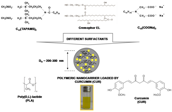
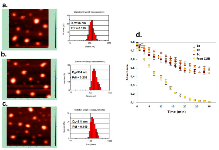
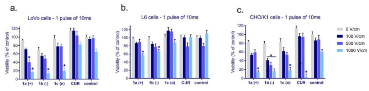
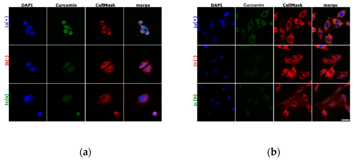
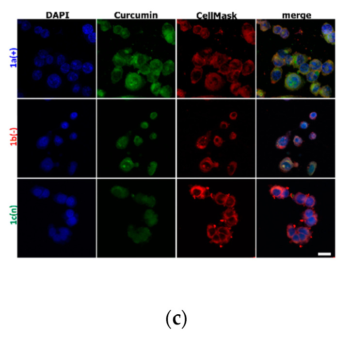
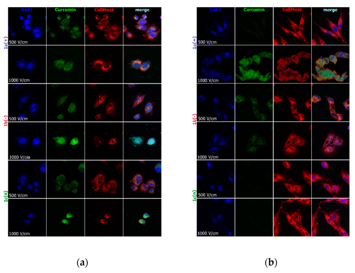
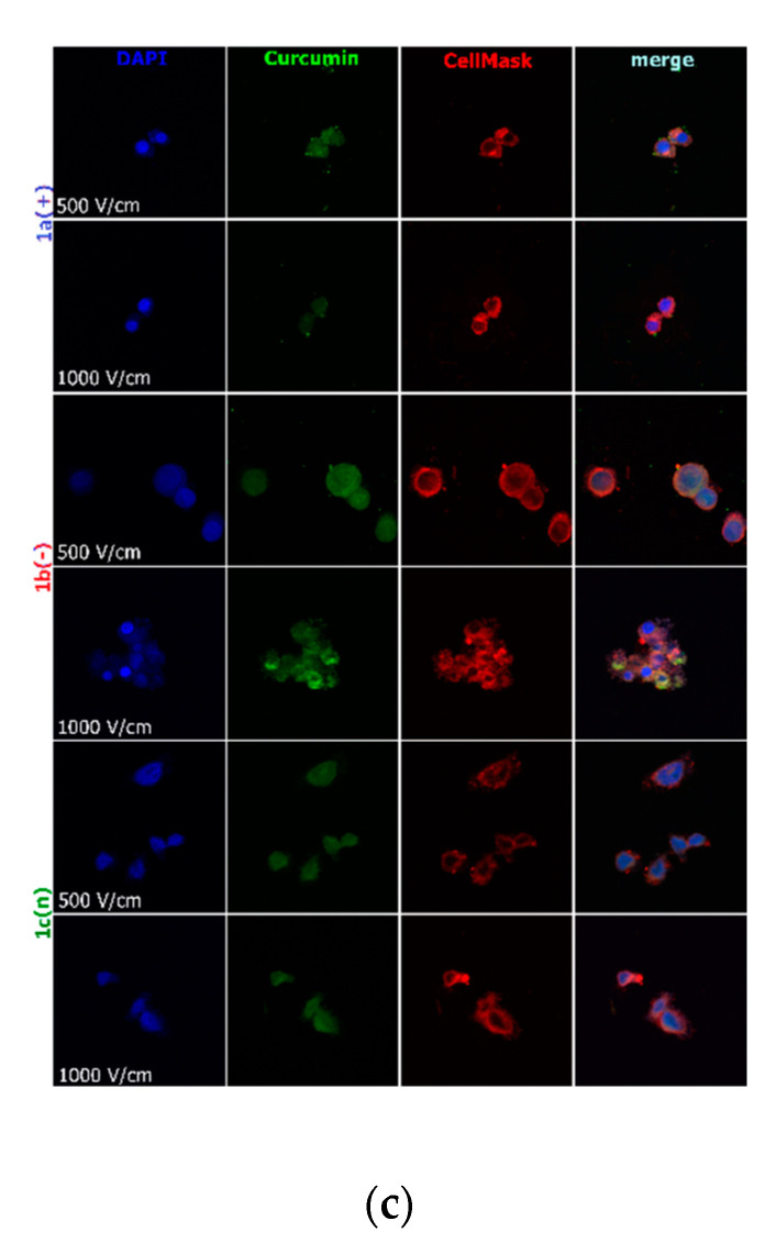
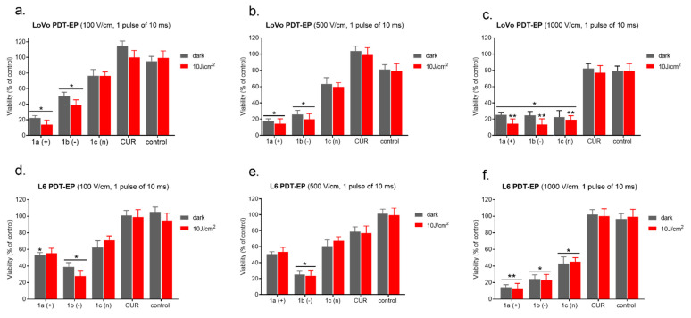
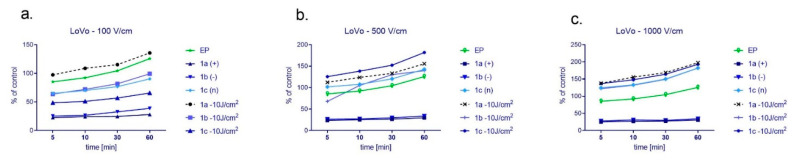
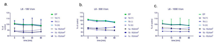
(1) Background: The size and surface charge are the most significant parameters of nanocarriers that determine their efficiency and potential application. The poor cell uptake of encapsulated drugs is the main limitation in anticancer treatment. The well-defined properties of nanocarriers will enable to target specific tissue and deliver an active cargo. (2) Methods: In the current study, poly(D,L -lactide) (PLA) nanocarriers loaded with curcumin (CUR) and differing surface charge were evaluated for transport efficacy in combination with electroporation (EP) in dependence on the type of cells. The obtained CUR-loaded nanoparticles with diameters ranging from 195 to 334 nm (derived from dynamic light scattering (DLS)) were characterized by atomic force microscopy (AFM) (morphology and shape) and Doppler electrophoresis (ζ-potential) as well as UV-vis spectroscopy (CUR encapsulation efficiency (about 90%) and photobleaching rate). The drug delivery properties of the obtained PLA nanocarriers enhanced by electroporation were assessed in human colon cancer cells (LoVo), excitable normal rat muscle cells (L6), and free of voltage-gated ion channels cells (CHO-K1). CLSM studies, viability, and ROS release were performed to determine the biological effects of nanocarriers. (3) Results: The highest photodynamic activity indicated anionic nanocarriers (1a) stabilized by C12(COONa)2 surfactant. Nanocarriers were cytotoxic for LoVo cells and less cytotoxic for normal cells. ROS release increased in cancer cells with the increasing electric field intensity, irradiation, and time after EP. Muscle L6 cells were less sensitive to electric pulses. (4) Conclusions: EP stimulation for CUR-PLA nanocarriers transport was considered to improve the regulated and more effective delivery of nanosystems differing in surface charge.

Keywords: PLA nanocarriers; colon cancer; curcumin; electroporation; surface charge.


Conflict of interest statement

The authors declare no conflict of interest.

Figures

Scheme 1
Scheme 1
The representation of the nanosystems composition.
Figure 1
Figure 1
Characteristics of CUR-loaded nanocarriers. AFM images and DLS size distribution graphs of CUR-loaded nanocarriers stabilized by C12(TAPAMS)2 (a), C12(COONa)2 (b), and Cremophor EL (c); photobleaching of 4 μM encapsulated and free CUR in aqueous solution during irradiation with visible light (100 mW/cm²) measured by UV-Vis absorption spectra (d). Points present the difference in the absorption at λmax = 440 nm for loaded nanocarriers and λmax = 430 nm for the native form of photosensitizer as a function of time.
Figure 2
Figure 2
The cytotoxicity of free curcumin and curcumin ([CUR] = 4 μM) loaded in nanosystems evaluated after 24 h in (a) colon cancer cells (LoVo); (b) skeletal muscle cells (L6); and (c) hamster ovarian fibroblasts (CHO/K1). * p < 0.05.
Figure 3
Figure 3
Cells incubated with encapsulated curcumin and stained for nuclei (DAPI) and cell membrane (CellMask DeepRed) visualization: (a) hamster ovarian fibroblasts (CHO-K1 cells); (b) normal rat skeletal muscle cells (L6 cells); (c) colon adenocarcinoma cells ( LoVo cells). The white scale bar corresponds to 10 µm. CUR concentration was 4 μM.
Figure 3
Figure 3
Cells incubated with encapsulated curcumin and stained for nuclei (DAPI) and cell membrane (CellMask DeepRed) visualization: (a) hamster ovarian fibroblasts (CHO-K1 cells); (b) normal rat skeletal muscle cells (L6 cells); (c) colon adenocarcinoma cells ( LoVo cells). The white scale bar corresponds to 10 µm. CUR concentration was 4 μM.
Figure 4
Figure 4
Cells electroporated (500 and 1000 V/cm) with encapsulated curcumin and stained for nuclei (DAPI) and cell membrane (CellMask DeepRed) visualization: (a) hamster ovarian fibroblasts (CHO-K1 cells); (b) normal rat skeletal muscle cells (L6 cells); (c) colon adenocarcinoma cells (LoVo cells). CUR concentration was 4 μM.
Figure 4
Figure 4
Cells electroporated (500 and 1000 V/cm) with encapsulated curcumin and stained for nuclei (DAPI) and cell membrane (CellMask DeepRed) visualization: (a) hamster ovarian fibroblasts (CHO-K1 cells); (b) normal rat skeletal muscle cells (L6 cells); (c) colon adenocarcinoma cells (LoVo cells). CUR concentration was 4 μM.
Figure 5
Figure 5
Photodynamic reaction after 10 min combined with EP in LoVo cells ((ac)—upper panel) and normal L6 cells ((df)—bottom panel). CUR concentration was 4 μM. * p ≤ 0.05, ** p ≤ 0.01.
Figure 6
Figure 6
The level of ROS in colon cancer LoVo cells after cyto- and photocytotoxic reaction supported by electroporation using the following electric field intensity (a) 100 V/cm, (b) 500 V/cm, and (c) 1000 V/cm. Results were normalized and presented as a percentage of untreated control cells. CUR concentration was 4 μM.
Figure 7
Figure 7
The level of ROS in normal L6 cells after cyto- and photocytotoxic reaction supported by electroporation using the following electric field intensity (a) 100 V/cm, (b) 500 V/cm, and (c) 1000 V/cm. Results were normalized and presented as a percentage of untreated control cells. CUR concentration was 4 μM.

Similar articles

Cited by

References

    1. Daraba O.M., Cadinoiu A.N., Rata D.M., Atanase L.I., Vochita G. Antitumoral drug-loaded biocompatible polymeric nanoparticles obtained by non-aqueous emulsion polymerization. Polymers. 2020;12:1018. doi: 10.3390/polym12051018. - DOI - PMC - PubMed
    1. Akhtar R., Chandel S., Sarotra P., Medhi B. Current status of pharmacological treatment of colorectal cancer. World J. Gastrointest. Oncol. 2014;6:177–183. doi: 10.4251/wjgo.v6.i6.177. - DOI - PMC - PubMed
    1. Mohelnikova-Duchonova B., Melichar B., Soucek P. FOLFOX/FOLFIRI pharmacogenetics: The call for a personalized approach in colorectal cancer therapy. World J. Gastroenterol. 2014;20:10316–10330. doi: 10.3748/wjg.v20.i30.10316. - DOI - PMC - PubMed
    1. Carethers J.M. Review: Systemic treatment of advanced colorectal cancer: Tailoring therapy to the tumor. Ther. Adv. Gastroenterol. 2008;1:33–42. doi: 10.1177/1756283X08093607. - DOI - PMC - PubMed
    1. Ruiz De Porras V., Bystrup S., Martínez-Cardús A., Pluvinet R., Sumoy L., Howells L., James M.I., Iwuji C., Manzano J.L., Layos L., et al. Curcumin mediates oxaliplatin-acquired resistance reversion in colorectal cancer cell lines through modulation of CXC-Chemokine/NF-κB signalling pathway. Sci. Rep. 2016;6:24675. doi: 10.1038/srep24675. - DOI - PMC - PubMed

LinkOut - more resources