. 2022 Jan 26;23(3):1387.
doi: 10.3390/ijms23031387.

Galangin Reverses H2O2-Induced Dermal Fibroblast Senescence via SIRT1-PGC-1α/Nrf2 Signaling

Affiliations

Galangin Reverses H2O2-Induced Dermal Fibroblast Senescence via SIRT1-PGC-1α/Nrf2 Signaling

Jian-Jr Lee et al. Int J Mol Sci. .

Abstract

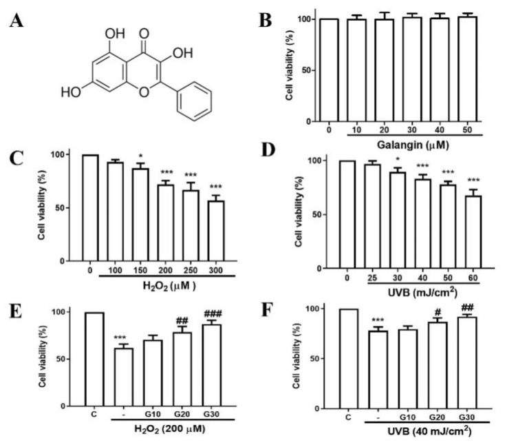
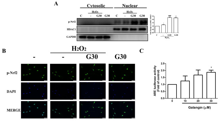
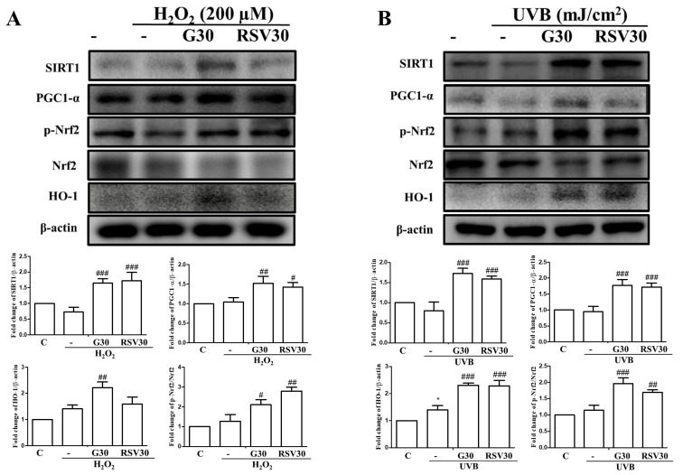
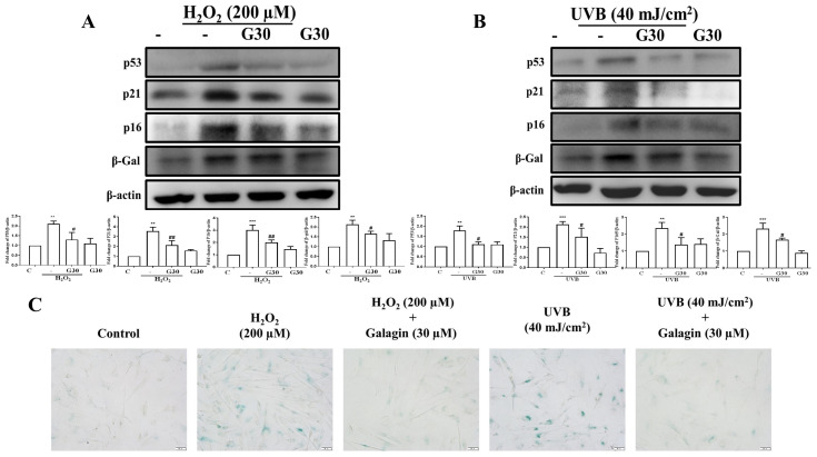
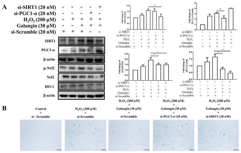
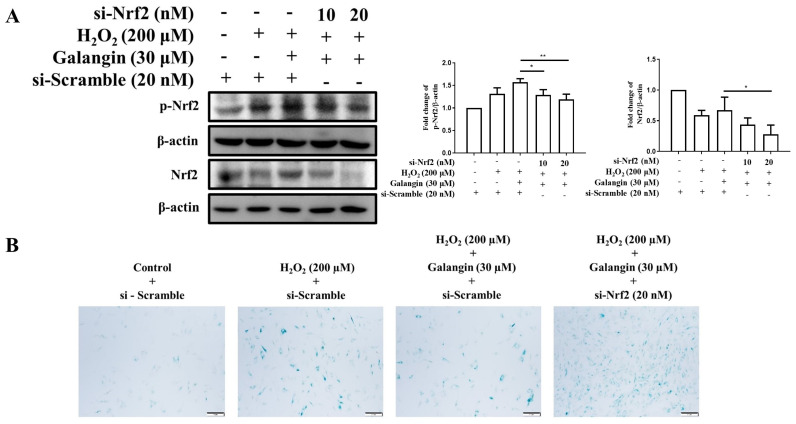
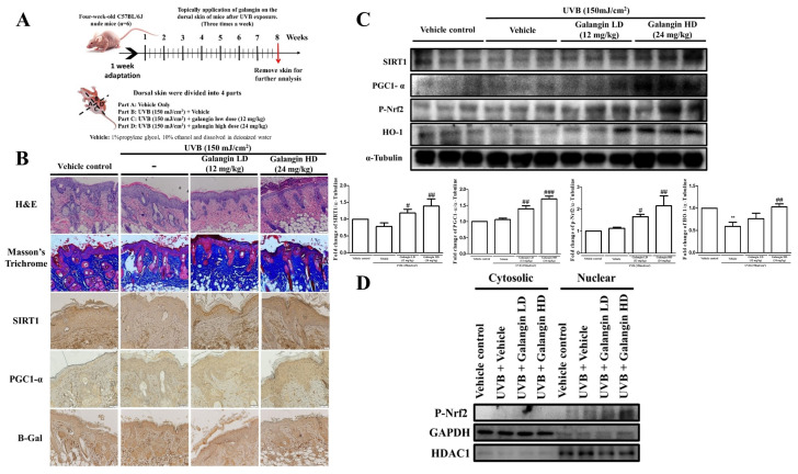
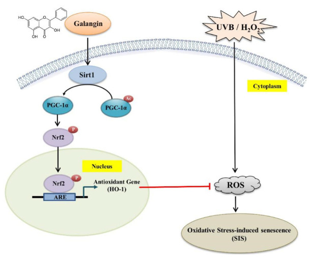
UV radiation and H2O2 are the primary factors that cause skin aging. Both trigger oxidative stress and cellular aging. It has been reported that deacetylase silent information regulator 1 (SIRT1), a longevity gene, enhances activation of NF-E2-related factor-2 (Nrf2), as well as its downstream key antioxidant gene hemeoxygenase-1 (HO-1), to protect cells against oxidative damage by deacetylating the transcription coactivator PPARγ coactivator-1α (PGC-1α). Galangin, a flavonoid, possesses anti-oxidative and anti-inflammatory potential. In the present study, we applied Ultraviolet B/H2O2-induced human dermal fibroblast damage as an in vitro model and UVB-induced photoaging of C57BL/6J nude mice as an in vivo model to investigate the underlying dermo-protective mechanisms of galangin. Our results indicated that galangin treatment attenuates H2O2/UVB-induced cell viability reduction, dermal aging, and SIRT1/PGC-1α/Nrf2 signaling activation. Furthermore, galangin treatment enhanced Nrf2 activation and nuclear accumulation, in addition to inhibiting Nrf2 degradation. Interestingly, upregulation of antioxidant response element luciferase activity following galangin treatment indicated the transcriptional activation of Nrf2. However, knockdown of SIRT1, PGC-1α, or Nrf2 by siRNA reversed the antioxidant and anti-aging effects of galangin. In vivo evidence further showed that galangin treatment, at doses of 12 and 24 mg/kg on the dorsal skin cells of nude mice resulted in considerably reduced UVB-induced epidermal hyperplasia and skin senescence, and promoted SIRT1/PGC-1α/Nrf2 signaling. Furthermore, enhanced nuclear localization of Nrf2 was observed in galangin-treated mice following UVB irradiation. In conclusion, our data indicated that galangin exerts anti-photoaging and antioxidant effects by promoting SIRT1/PGC-1α/Nrf2 signaling. Therefore, galangin is a potentially promising agent for cosmetic skin care products against UV-induced skin aging.

Keywords: HS68 human dermal fibroblast cells; PGC-1α; SIRT1; UVB; senescence.


Conflict of interest statement

The authors declare no conflict of interest.

Figures

Figure 1
Figure 1
Galangin inhibits UVB- and H2O2-induced proliferation reduction in HS68 cells. (A) Structure of galangin (3,5,7-trihydroxyflavone). (BD) HS68 cells were treated with galangin, H2O2, and UVB, at the indicated concentrations/intensities for 24 h. (E,F) HS68 cells were exposed to H2O2 (200 μM) or UVB (40 mJ/cm2) and then co-treated with galangin at various concentrations (10–30 μM). The MTT assay was used to determine cell viability. Values shown are means ± SE. Quantification of the results is shown (n = 3) * p < 0.05, *** p < 0.001 vs. control cells; # p < 0.05, ## p < 0.01, ### p < 0.001 vs. H2O2- or UVB-treated cells.
Figure 2
Figure 2
Effects of galangin on the Sirt1/PGC-1α/Nrf2 pathway and upregulation of antioxidant genes (HO-1) in HS68 cells exposed to UVB/H2O2. (A) HS68 cells were treated with different concentrations of galangin for 24 h. (B) The binding of Ub with Nrf2 was detected using immunoprecipitation under different concentrations of galangin for 24 h. (C,D) HS68 cells were exposed to H2O2 (200 μM) or UVB (40 mJ/cm2) and then co-treated with galangin at a specific concentration (30 μM). Protein levels of Sirt1, PGC-1α, Nrf2, p-Nrf2, and HO-1 were detected by Western blot. Values shown are means ± SE. Quantification of the results is shown (n = 3) * p < 0.05, ** p < 0.01, *** p < 0.001 vs. untreated control cells; # p < 0.05, ### p < 0.001 vs. H2O2/UVB-treated cells.
Figure 3
Figure 3
Galangin triggers Nrf2 nuclear translocation and ARE transcriptional activation in HS68 cells exposed to H2O2. (A,B) HS68 cells were exposed to H2O2 (200 μM) or UVB (40 mJ/cm2) and then co-treated with galangin at a specific concentration (30 μM). p-Nrf2 expression was estimated in the cytosolic and nuclear fractions. The p-Nrf2 protein level was detected by Western blot. (C) p-Nrf2 nuclear translocation was further ascertained by immunofluorescence staining. An anti-Nrf2 antibody and a FITC-conjugated second antibody were used to detect p-Nrf2 cellular distribution. DAPI staining indicated the nucleus location (blue). The images were acquired using florescence microscopy (200×). HS68 cells were transfected with ARE-luciferase construct for 24 h. The cells were then treated with different doses of galangin (10, 20, and 30 µM) for 24 h and assayed for luciferase activity. Values shown are means ± SE. Quantification of the results is shown (n = 3) * p < 0.05, *** p < 0.001 vs. untreated control cells; ### p < 0.001 vs. H2O2-treated cells.
Figure 4
Figure 4
Galangin enhances the Sirt1/PGC-1α/Nrf2 pathway and its downstream gene (HO-1), as well as resveratrol (Sirt1 activator), in HS68 cells under UVB/H2O2-induced damage. (A,B) HS68 cells were exposed to H2O2 (200 μM) or UVB (40 mJ/cm2) and then co-treated with galangin (30 μM) or resveratrol (30 µM) as indicated in related graphs. The protein levels of Sirt1, PGC-1α, p-Nrf2, Nrf2, and HO-1 were detected by Western blot. Values shown are means ± SE. Quantification of the results is shown (n = 3) * p < 0.05 vs. untreated control cells; # p < 0.05, ## p < 0.01, ### p < 0.001 vs. H2O2/UVB-treated cells.
Figure 5
Figure 5
Anti-aging effects of galangin on UVB/H2O2-induced senescence of HS68 cells. (A,B) HS68 cells were exposed to H2O2 (200 μM) or UVB (40 mJ/cm2) and then co-treated with galangin at a specific concentration (30 μM). Protein levels of p53, p21, p16, and β-Gal were detected by Western blot. (C) After treatment, cells were stained using an SA-β-Gal staining kit to detect SA-β-Gal activity. Positive cells were stained in green color, which indicated senescent cells. Values shown are means ± SE. Quantification of the results is shown (n = 3) ** p < 0.01, *** p < 0.001 vs. untreated control cells; # p < 0.05, ## p < 0.01, vs. H2O2/UVB-treated cells.
Figure 6
Figure 6
Silencing of Sirt1 or PGC-1α diminishes the protective effects of galangin under H2O2 exposure in HS68 cells. (A) Levels of Nrf2, p-Nrf2, and HO-1 measured by Western blot in response to Sirt1 and PGC-1α silencing. (B) After treatment, cells were stained using an SA-β-Gal staining kit to detect SA-β-Gal activity. Positive cells were stained in green, which indicates senescent cells. Values shown are means ± SE. Quantification of the results is shown (n = 3) * p < 0.05, ** p < 0.01, vs. galangin plus H2O2-treated cells.
Figure 7
Figure 7
Silencing of Nrf2 diminishes the protective effects of galangin under H2O2 exposure in HS68 cells. (A) p-Nrf (active) and Nrf protein levels were detected by Western blotting in response to siRNA silencing to ensure knockdown efficiency. β-actin acted as loading control. (B) After treatment, cells were stained using an SA-β-Gal staining kit to detect SA-β-Gal activity. Positive cells were stained in green, which indicates senescent cells. Values shown are means ± SE. Quantification of the results is shown (n = 3) * p < 0.05, ** p < 0.01, vs. galangin plus H2O2-treated cells.
Figure 8
Figure 8
Galangin alleviates UVB-induced skin photodamage in C57BL/6J nude mice. (A) The experimental animal protocol has been described in detail in the Materials and Methods section. (B) Skin morphology and collagen levels were determined by H&E stain and Masson’s trichrome stain, respectively. Sirt1, PGC1-α, and β-Gal were detected by immunohistochemistry. (C) Western blot analysis of SIRT1, PGC1-α, p-Nrf, and HO-1 protein levels in the dorsal skin of the mice. α-Tubulin acted as a loading control. (D) p-Nrf2 expression in the cytosol and nucleus was analyzed using a nucleus and cytosol extraction kit and detected by Western blotting. Values shown are means ± SE. Quantification of the results is shown (n = 3) ** p < 0.01 vs. vehicle control group; # p < 0.05, ## p < 0.01, ### p < 0.001 vs. UVB-irradiated group.
Figure 9
Figure 9
Scheme summarizing the inhibition of H2O2-induced skin oxidative damage by galangin via the upregulation of antioxidant genes (HO-1) through the Sirt1/PGC-1α/Nrf2 pathway in HS68 human dermal fibroblast cells. Sirt 1: silent information regulator 1; PGC-1α: PPARγ coactivator-1α; Nrf2: NF-E2-related factor-2; HO-1: hemeoxygenase-1.

Similar articles

Cited by

References

    1. Ganceviciene R., Liakou A.I., Theodoridis A., Makrantonaki E., Zouboulis C.C. Skin anti-aging strategies. Dermatoendocrinology. 2012;4:308–319. doi: 10.4161/derm.22804. - DOI - PMC - PubMed
    1. Addor F.A.S. Beyond photoaging: Additional factors involved in the process of skin aging. Clin. Cosmet. Investig. Dermatol. 2018;11:437–443. doi: 10.2147/CCID.S177448. - DOI - PMC - PubMed
    1. Schieber M., Chandel N.S. ROS function in redox signaling and oxidative stress. Curr. Biol. 2014;24:R453–R462. doi: 10.1016/j.cub.2014.03.034. - DOI - PMC - PubMed
    1. Wang A.S., Dreesen O. Biomarkers of Cellular Senescence and Skin Aging. Front. Genet. 2018;9:247. doi: 10.3389/fgene.2018.00247. - DOI - PMC - PubMed
    1. Ho C.C., Ng S.C., Chuang H.L., Wen S.Y., Kuo C.H., Mahalakshmi B., Huang C.Y., Kuo W.W. Extracts of Jasminum sambac flowers fermented by Lactobacillus rhamnosus inhibit H2O2—And UVB-induced aging in human dermal fibroblasts. Environ. Toxicol. 2021;36:607–619. doi: 10.1002/tox.23065. - DOI - PubMed

MeSH terms

LinkOut - more resources