. 2022 Jan 26;23(3):1401.
doi: 10.3390/ijms23031401.

Metabolites of Cannabis Induce Cardiac Toxicity and Morphological Alterations in Cardiac Myocytes

Affiliations

Metabolites of Cannabis Induce Cardiac Toxicity and Morphological Alterations in Cardiac Myocytes

Ayse Orme Merve et al. Int J Mol Sci. .

Abstract

Cannabis is one of the most commonly used recreational drugs worldwide. Rrecent epidemiology studies have linked increased cardiac complications to cannabis use. However, this literature is predominantly based on case incidents and post-mortem investigations. This study elucidates the molecular mechanism of Δ9-tetrahydrocannabinol (THC), and its primary metabolites 11-Hydroxy-Δ9-THC (THC-OH) and 11-nor-9-carboxy-Δ⁹-tetrahydrocannabinol (THC-COOH). Treatment of cardiac myocytes with THC-OH and THC-COOH increased cell migration and proliferation (p < 0.05), with no effect on cell adhesion, with higher doses (250-100 ng/mL) resulting in increased cell death and significant deterioration in cellular architecture. Conversely, no changes in cell morphology or viability were observed in response to THC. Expression of key ECM proteins α-SMA and collagen were up-regulated in response to THC-OH and THC-COOH treatments with concomitant modulation of PI3K and MAPK signalling. Investigations in the planarian animal model Polycelis nigra demonstrated that treatments with cannabinoid metabolites resulted in increased protein deposition at transection sites while higher doses resulted in significant lethality and decline in regeneration. These results highlight that the key metabolites of cannabis elicit toxic effects independent of the parent and psychoactive compound, with implications for cardiotoxicity relating to hypertrophy and fibrogenesis.

Keywords: THC; cannabis; cardiac myocytes; cardiac toxicity; cytoskeleton; planaria.


Conflict of interest statement

The authors declare no conflict of interest.

Figures
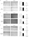
Figure 1
Figure 1
Metabolites of cannabis induce increased wound closure (A) H9c2 cardiomyocytes were exposed to 100 ng/mL of THC for 24 h, (B) 250 ng/mL of THC-OH for 24 h and (C) 250 ng/mL of THC-COOH for 24 h compared with complete growth medium control following scratch wound treatments. The number of migrant cells were quantified by ImageJ analysis software and compared against untreated control cells. The migratory distance of cardiomyocytes in terms of wound healing was determined manually by equivalence of the scale bar and compared with untreated control cells (scale bar was 1000 µm). Statistical comparison for treated vs. control was performed by a two-tailed Students unpaired t-test (* p < 0.05). n.s. refers to not statistically significant. Results represent the mean of three individual experiments (N = 3).
Figure 2
Figure 2
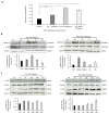
H9c2 changes in cell proliferation in response to cannabinoids. (A) H9c2 cardiomyocyte proliferation in response to THC, THC-OH and THC-COOH, represented data of five individual experiments. (B) Immunoblot analysis of ERK1/2 phosphorylation following THC-OH and THC-COOH (both 250 ng/mL) exposure. (C) Immunoblot analysis of AKT phosphorylation following THC-OH and THC-COOH (both 250 ng/mL) exposure. (B,C) Representative immunoblots of three independent experiments. Densitometry indicating relative band expression for each blot measured using ImageJ software. Statistical comparison for treated vs. control was performed by a two-tailed Students unpaired t-test (n.s. refers to not statistically significant, * p < 0.05, ** p < 0.01 and *** p < 0.001) for individual time points.
Figure 3
Figure 3
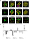
In-silico studies of THC and its metabolites THC-OH and THC-COOH modelled against key signalling regulators. (A) Interaction between ligands THC, THC-OH and THC-COOH with the ATP binding site known regulators; CB1, LIMK, ROCK, LRP5/6 and compared to CB1 as a positive control of a binding site that is permissive to cannabinoid activation. (B) Binding energies measured in kcal/mol generated by in-silico studies of THC and its metabolites THC-OH and THC-COOH modelled against key signalling regulators. (A,B) generated using the Scigress docking software.
Figure 4
Figure 4
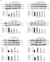
Cannabinoids differentially modulate ABP and ECM proteins. (A) Immunoblot analysis of Cofilin phosphorylation following THC-OH and THC-COOH exposure in H9c2 cardiomyocytes. (B) Time dependent protein detection of β-catenin following THC-OH and THC-COOH. (C) Western blot analysis of α-SMA and Col1α1 following THC-OH and THC-COOH. (AC) Representative immunoblots of three independent experiments. Drug treatment concentration for both THC-OH and THC-COOH were 250 ng/mL. Densitometry indicating relative band expression for each blot measured using ImageJ software. Statistical comparison for treated vs. control was performed by a two-tailed Students unpaired t-test (* p < 0.05, ** p < 0.01 and *** p < 0.001) for individual time points.
Figure 5
Figure 5
Dose dependent cannabinoid treatments result in morphological alterations of H9c2 cardiomyocytes. (AD) In response to both low dose and high dose THC treatments, 100–200 ng/mL, normal H9c2 cardiomyocytes morphology was retained with intercellular connections and spindle-like spreading present at 24 h following culture. (EH) Morphological alterations induced in H9c2 cardiomyocytes following exposure to 250 ng/mL THC-OH or THC-COOH. H9c2 cardiomyocytes indicated features of altered microstructural architecture, with membrane polarization, cell-substrate adhesion and retraction observed posterior to sites of membrane folding. Scale bar for each panel are presented below the respective image with numerical assignment indicating feature specific responses.
Figure 5
Figure 5
Dose dependent cannabinoid treatments result in morphological alterations of H9c2 cardiomyocytes. (AD) In response to both low dose and high dose THC treatments, 100–200 ng/mL, normal H9c2 cardiomyocytes morphology was retained with intercellular connections and spindle-like spreading present at 24 h following culture. (EH) Morphological alterations induced in H9c2 cardiomyocytes following exposure to 250 ng/mL THC-OH or THC-COOH. H9c2 cardiomyocytes indicated features of altered microstructural architecture, with membrane polarization, cell-substrate adhesion and retraction observed posterior to sites of membrane folding. Scale bar for each panel are presented below the respective image with numerical assignment indicating feature specific responses.
Figure 6
Figure 6
Low dose cannabinoids modulate regenerative capacity of Polycelis nigra. The effects of cannabinoids at low doses on the planarian flatworm regenerative capacity was assessed over 7 days. Decapitated planaria were treated with THC (100 ng/mL), THC-OH and THC-COOH (both 250 ng/mL) for one week after decapitation Arrows show increased regenerating areas.
Figure 7
Figure 7
High dose cannabinoids treatments induce toxicity in Polycelis nigra. (A) High dose drug concentration treatment of THC (1000 ng/mL), and its metabolites (both 2500 ng/mL) in decapitated planaria observed for a week for regeneration and toxicity. (B) THC-OH and THC-COOH demonstrated toxicity in high dose, while THC-OH caused rapid disintegration and THC-COOH showed slower disintegration in which structural integrity was protected for a longer period of time. The arrows highlight the slower disintergration seen in THC-COOH treatments versus that of TH-OH.

Similar articles

Cited by

References

    1. Knuth M., Temme O., Daldrup T., Pawlik E. Analysis of cocaine adulterants in human brain in cases of drug-related death. Forensic Sci. Int. 2018;285:86–92. doi: 10.1016/j.forsciint.2018.02.001. - DOI - PubMed
    1. Mittleman M.A., Mintzer D., Maclure M., Tofler G.H., Sherwood J.B., Muller J.E. Triggering of Myocardial Infarction by Cocaine. Circulation. 1999;99:2737–2741. doi: 10.1161/01.CIR.99.21.2737. - DOI - PubMed
    1. Egred M., Viswanathan G., Davis G.K. Myocardial infarction in young adults. Postgrad. Med. J. 2005;81:741–745. doi: 10.1136/pgmj.2004.027532. - DOI - PMC - PubMed
    1. Maceira A.M., Ripoll C., Cosin-Sales J., Igual B., Gavilan M., Salazar J., Belloch V., Pennell D.J. Long term effects of cocaine on the heart assessed by cardiovascular magnetic resonance at 3T. J. Cardiovasc. Magn. Reson. 2014;16:26. doi: 10.1186/1532-429X-16-26. - DOI - PMC - PubMed
    1. Wang Z.-J., Martin J.A., Gancarz A.M., Adank D.N., Sim F.J., Dietz D.M. Activin A is increased in the nucleus accumbens following a cocaine binge. Sci. Rep. 2017;7:43658. doi: 10.1038/srep43658. - DOI - PMC - PubMed

LinkOut - more resources