Review
. 2022 Jan 27;23(3):1455.
doi: 10.3390/ijms23031455.

The Discovery and Function of Filaggrin

Affiliations
Review

The Discovery and Function of Filaggrin

J Kenneth Hoober et al. Int J Mol Sci. .

Abstract

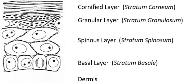
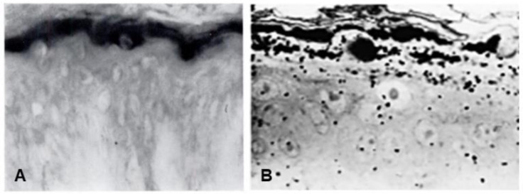
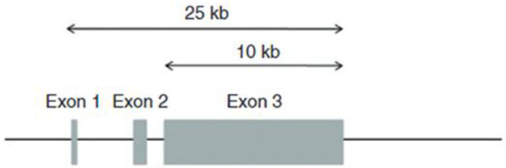
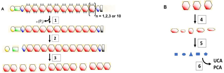
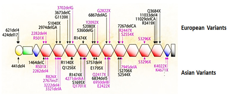
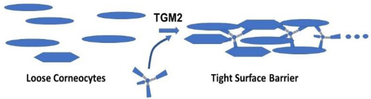
Keratohyalin granules were discovered in the mid-19th century in cells that terminally differentiate to form the outer, cornified layer of the epidermis. The first indications of the composition of these structures emerged in the 1960s from a histochemical stain for histidine, followed by radioautographic evidence of a high incidence of histidine incorporation into newly synthesized proteins in cells containing the granules. Research during the next three decades revealed the structure and function of a major protein in these granules, which was initially called the 'histidine-rich protein'. Steinert and Dale named the protein 'filaggrin' in 1981 because of its ability to aggregate keratin intermediate filaments. The human gene for the precursor, 'profilaggrin,' was reported in 1991 to encode 10, 11 or 12 nearly identical repeats. Remarkably, the mouse and rat genes encode up to 20 repeats. The lifetime of filaggrin is the time required for keratinocytes in the granular layer to move into the inner cornified layer. During this transition, filaggrin facilitates the collapse of corneocytes into 'building blocks' that become an impermeable surface barrier. The subsequent degradation of filaggrin is as remarkable as its synthesis, and the end-products aid in maintaining moisture in the cornified layer. It was apparent that ichthyosis vulgaris and atopic dermatitis were associated with the absence of this protein. McLean's team in 2006 identified the cause of these diseases by discovering loss-of-function mutations in the profilaggrin gene, which led to dysfunction of the surface barrier. This story illustrates the complexity in maintaining a healthy, functional epidermis.

Keywords: atopic dermatitis; corneodesmosomes; eczema; filaggrin; histidine-rich protein; ichthyosis vulgaris; keratohyalin granules; loss-of-function mutations; profilaggrin; transglutaminase.


Conflict of interest statement

J.K.H. and L.L.E. declare that they are cofounders of Susavion Biosciences, Inc., in which they hold shares.

Figures

Figure 1
Figure 1
A sketch of the epidermis of the newborn rat [4].
Figure 2
Figure 2
(A) Human skin stained with the Pauly reagent for histidine [5]. (B) Light microscopic radioautograph of newborn rat epidermis 15 min after intraperitoneal injection of [3H]histidine [12].
Figure 3
Figure 3
The human profilaggrin gene on chromosome 1q21. Exon 2 contains the translation initiation codon. The entire coding region of the protein is within the uninterrupted exon 3.
Figure 4
Figure 4
Flowchart of the processing of profilaggrin to free amino acids. (A). Proflaggrin to filaggrin. (1) Phospho-profilaggrin is dephosphorylated by phosphatases. (2) The A (yellow) and B (green) domains are cleaved from profilaggrin by furin, PACE4 (Paired basic Amino acid Cleaving Enzyme 4) and endoproteinase-1 (PEP1) [50,51]. (3) Linker sequences of human profilaggrin (FLYQVST) are cleaved by skin-specific retrovirus-like aspartic protease (SASPase) [52], channel-activating serine protease (CAP1) [53] and matriptase (MT-SP1) [54] to monomeric filaggrin. Aminopeptidases and carboxypeptidases are likely involved in trimming the new termini [37]. (B). Filaggrin to NMF. (4) Arginine residues in filaggrin and keratin are converted to citrulline by peptidylarginine deiminase (PAD1 or 3) [49,55]. Deiminated filaggrin is cleaved by calpain-1 and caspase-14 (at VSQD and HSED sequences) to filaggrin fragments [55,56,57]. (5) Filaggrin fragments are digested by neutral cysteine protease (bleomycin hydrolase) to amino acids [57]. Aminopeptidases and carboxypeptidases are also likely involved. (6) Histidine is converted by histidine deaminase to trans-urocanic acid (UCA), which has a UV spectrum similar to that of nucleic acids and proteins [58] and provides a natural sunscreen [59]. Glutamine is non-enzymatically converted to 2-pyrrolidone carboxylic acid (PCA). These hydrophilic final products contribute to the moisturizing of the skin [56,57,60,61]. Kezic et al. [62] and de Veer et al. [63] provided excellent reviews of proteolytic processing of filaggrin and differentiation of corneocytes.
Figure 5
Figure 5
Filaggrin mutations among populations. Mutations that are recurrent in European and Asian populations are indicated in red, and rare or family specific mutations are in black. These mutations are either nonsense mutations or out-of-frame insertions or deletions that are predicted to cause loss-of-function. Exon 3 of the gene contains the complete sequence for profilaggrin, shown here with 10 repeats as orange hexagons. The yellow circle is the S-100-like Ca2+-binding domain, the green rectangle is the B domain, and the blue diamonds are incomplete repeats [65] (with permission from A. D. Irvine).
Figure 6
Figure 6
Number of publications that refer to filaggrin as a function of year.
Figure 7
Figure 7
Corneodesmosomes. (A) Filaggrin-deficient (FLG−/−) corneocytes from tape-stripped stratum corneum of patients with AD express corneodesmosin on villus-like projections that cover the entire cell surface. (B) In contrast, corneocytes from the tightly packed stratum corneum of wild-type (FLG+/+) patients contain corneodesmosin mostly on lateral rims of the cells. The images were prepared by incubating corneocytes with anti-corneodesmosin antibodies followed by immunogold labeling and scanning electron microscopy [124] (with permission from S. Kezic).
Figure 8
Figure 8
Model for the role of a tetravalent peptide in facilitating cross-linking of corneocytes by TGM2.

References

    1. Holbrook K.A. Biologic structure and function: Perspectives on morphologic approaches to the study of the granular layer keratinocyte. J. Investig. Dermatol. 1989;92:s84–s104. doi: 10.1038/jid.1989.36. - DOI - PubMed
    1. Rothman S. Keratinization in historical perspective. In: Montagna W., Lobitz W.C. Jr., editors. The Epidermis. Academic Press; New York, NY, USA: 1964. pp. 1–14.
    1. Burgdorf W.H.C., Bickers D.R. The scientific legacy of Stephen Rothman. J. Investig. Dermatol. 2015;135:954–959. doi: 10.1038/jid.2014.447. - DOI - PMC - PubMed
    1. Hoober J.K. Ph.D. Thesis. University of Michigan; Ann Arbor, MI, USA: 1965. The Differential Incorporation of Amino Acids (In Vivo) into Proteins of the Newborn Rat Epidermis.
    1. Reaven E.P., Cox A.J., Jr. The histochemical localization of histidine in the human epidermis and its relationship to zinc binding. J. Histochem. Cytochem. 1963;11:782–790. doi: 10.1177/11.6.782. - DOI

LinkOut - more resources