. 2022 Jan 27;23(3):1476.
doi: 10.3390/ijms23031476.

ARPC1B Is Associated with Lethal Prostate Cancer and Its Inhibition Decreases Cell Invasion and Migration In Vitro

Affiliations

ARPC1B Is Associated with Lethal Prostate Cancer and Its Inhibition Decreases Cell Invasion and Migration In Vitro

Yaser Gamallat et al. Int J Mol Sci. .

Abstract

ARPC1B (Actin Related Protein 2/3 Complex Subunit 1B) has been found to be involved in platelet abnormalities of immune-mediated inflammatory disease and eosinophilia. However, its role in prostate cancer (PCa) has not been established. We characterized the role of ARPC1B in PCa invasion and metastasis and investigated its prognosis using in vitro cellular models and PCa clinical data. Higher immunohistochemistry (IHC) expressions of ARPC1B were observed in localized and castrate resistant PCa (CRPC) vs. benign prostate tissue (p < 0.01). Additionally, 47% of patients with grade group 5 (GG) showed high ARPC1B expression vs. other GG patients. Assessing ARPC1B expression in association with two of the common genetic aberrations in PCa (ERG and PTEN) showed significant association to overall and cause-specific survival for combined assessment of ARPC1B and PTEN, and ARPC1B and ERG. Knockdown of ARPC1B impaired the migration and invasion of PC3 and DU145 PCa cells via downregulation of Aurora A kinase (AURKA) and resulted in the arrest of the cells in the G2/M checkpoint of the cell cycle. Additionally, higher ARPC1B expression was observed in stable PC3-ERG cells compared to normal PC3, supporting the association between ERG and ARPC1B. Our findings implicate the role of ARPC1B in PCa invasion and metastasis in association with ERG and further support its prognostic value as a biomarker in association with ERG and PTEN in identifying aggressive phenotypes of PCa cancer.

Keywords: ARPC1B; ERG; PTEN; immigration; invasion; prognosis; prostate cancer.


Conflict of interest statement

The authors declare no conflict of interest.

Figures

Figure 1
Figure 1
ARPC1B expression in clinical prostate cancer cases. Immunohistochemistry staining (IHC) shows ARPC1B expression in Benign (BEN, 10×), localized prostate cancer (PCA, 4×), and castration resistance prostate cancer (CRPC, 10×) in human tissue samples. The mean expression of ARPC1B in BEN, PCA, and CRPC in human tissue samples. ARPC1B protein expression levels were scored through IHC. Each sample was scored semi quantitatively using four-tiered system (negative—0; weak—1; moderate—2; strong—3). The error bars indicate the Standard error of the mean. Student t-test was performed, p value < 0.05 was considered significant between prostate cancer CRPC, and localized PCa was compared with benign. p < 0.0144, p < 0.0123.
Figure 2
Figure 2
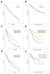
Kaplan–Meier (KM) curves demonstrating ARPC1B expression in relation to prostate cancer cases overall survival and cause-specific survival. (A) overall survival according to ARPC1B IHC score of and (B) KM curves for cause-specific survival with ARPC1B score (C,D) ARPC1B and PTEN loss. (E,F) ARPC1B and ERG gain. Each sample was scored semi-quantitatively for ARPC1B using four-tiered system (negative—0; weak—1; moderate—2; strong—3). PTEN loss = PTEN negative staining. PTEN gain = weak, moderate, or high staining.
Figure 3
Figure 3
ARPC1B genomic alteration KM for (A) PCa overall survival and (B) Bladder cancer progression free survival. Data obtained from cBioPortal (contains all TCGA datasets, ~12.5 K samples combined for the bladder and prostate cancer together.
Figure 4
Figure 4
Expression of ARPC1B in PCa cell lines. (A) Western blot analysis of ARPC1B expression in PCa cell lines: RWPE1, PC3, C4-2, DU145, LNCaP, and VCAP. (B) Knockdown of PC3 and DU145 cell lines using ARPC1B siRNA#1, siRNA#2, or control scramble siRNA as the negative control. (C) The expression of ARPC1B in PC3-ERG overexpression cells (D) Western blot analysis of ARPC1B knockdown in PC3-ERG cells using ARPC1B, siRNA#1, or scramble siRNA as the negative control.
Figure 5
Figure 5
Knockdown of ARPC1B in PC3 cell line attenuates PCa cell proliferation and migration. (A) Relative PC3 cells proliferation using MTS assay over three days. Statistical significance calculated using, two-way ANOVA, *** shows p < 0.001. Error bars indicate the SEM. (B) wound healing assay for PC3 cells after ARPC1B knockdown at 0, 24, and 48 h.
Figure 6
Figure 6
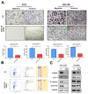
ARPC1 knockdown attenuates PCa cell migration and invasion. (A) Migration and invasion assay for PC3 and DU145 cell lines after 48 h of ARPC1B knockdown. *** p < 0.001, ** p < 0.01. Scale bar 400 µM. (B) Cell cycle analysis using PI staining. Data analyzed and presented using FlowJo. (C) Western blot analysis of AURKA, Lamin A, N-Cadherin, Cyclin B1 in PC3, and PC3-ERG cell transfected with either ARPC1B, siRNA, or control scramble siRNA as the negative control. GAPDH used as loading control.

Similar articles

Cited by

References

    1. Siegel R.L., Miller K.D., Fuchs H.E., Jemal A. Cancer Statistics, 2021. CA Cancer J. Clin. 2021;71:7–33. doi: 10.3322/caac.21654. - DOI - PubMed
    1. Kumagai K., Nimura Y., Mizota A., Miyahara N., Aoki M., Furusawa Y., Takiguchi M., Yamamoto S., Seki N. Arpc1b gene is a candidate prediction marker for choroidal malignant melanomas sensitive to radiotherapy. Investig. Ophthalmol. Vis. Sci. 2006;47:2300–2304. doi: 10.1167/iovs.05-0810. - DOI - PubMed
    1. Randzavola L.O., Strege K., Juzans M., Asano Y., Stinchcombe J.C., Gawden-Bone C.M., Seaman M.N., Kuijpers T.W., Griffiths G.M. Loss of ARPC1B impairs cytotoxic T lymphocyte maintenance and cytolytic activity. J. Clin. Investig. 2019;129:5600–5614. doi: 10.1172/JCI129388. - DOI - PMC - PubMed
    1. Goley E.D., Welch M.D. The ARP2/3 complex: An actin nucleator comes of age. Nat. Rev. Mol. Cell Biol. 2006;7:713–726. doi: 10.1038/nrm2026. - DOI - PubMed
    1. Welch M.D., DePace A.H., Verma S., Iwamatsu A., Mitchison T.J. The human Arp2/3 complex is composed of evolutionarily conserved subunits and is localized to cellular regions of dynamic actin filament assembly. J. Cell Biol. 1997;138:375–384. doi: 10.1083/jcb.138.2.375. - DOI - PMC - PubMed

Substances