Review
. 2022 Jan 27;23(3):1454.
doi: 10.3390/ijms23031454.

Allosterism in the PDZ Family

Affiliations
Review

Allosterism in the PDZ Family

Amy O Stevens et al. Int J Mol Sci. .

Abstract

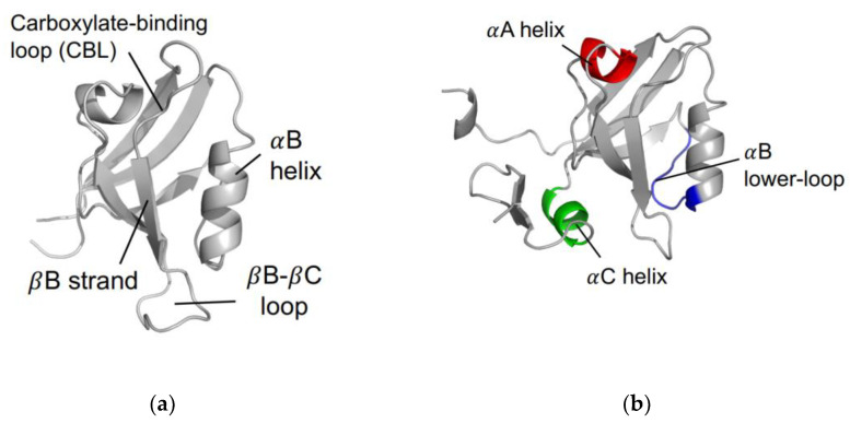
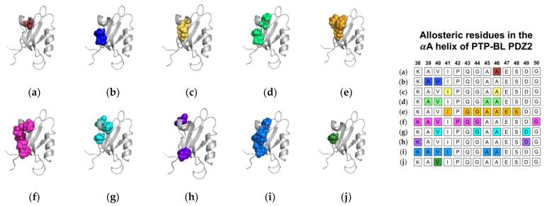
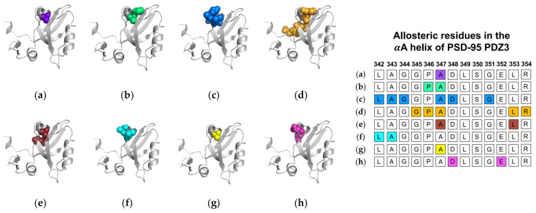
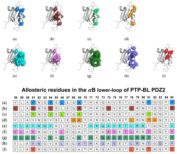
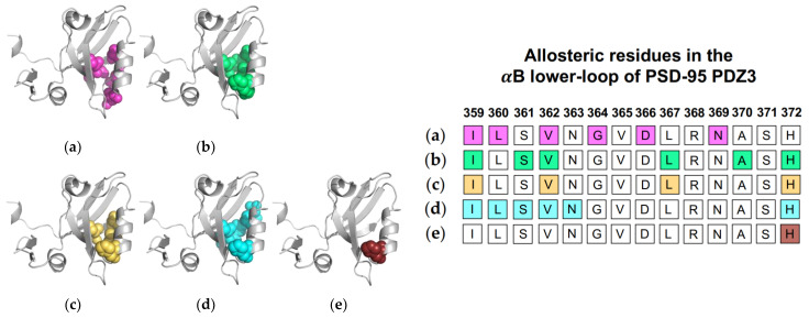
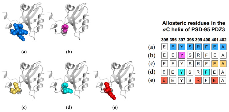
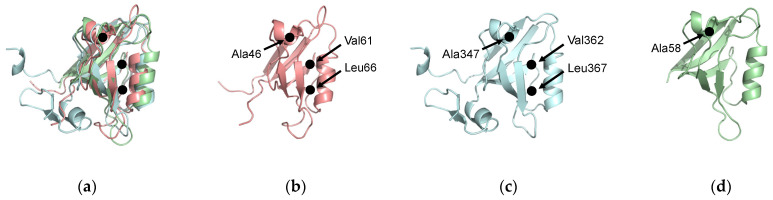
Dynamic allosterism allows the propagation of signal throughout a protein. The PDZ (PSD-95/Dlg1/ZO-1) family has been named as a classic example of dynamic allostery in small modular domains. While the PDZ family consists of more than 200 domains, previous efforts have primarily focused on a few well-studied PDZ domains, including PTP-BL PDZ2, PSD-95 PDZ3, and Par6 PDZ. Taken together, experimental and computational studies have identified regions of these domains that are dynamically coupled to ligand binding. These regions include the αA helix, the αB lower-loop, and the αC helix. In this review, we summarize the specific residues on the αA helix, the αB lower-loop, and the αC helix of PTP-BL PDZ2, PSD-95 PDZ3, and Par6 PDZ that have been identified as participants in dynamic allostery by either experimental or computational approaches. This review can serve as an index for researchers to look back on the previously identified allostery in the PDZ family. Interestingly, our summary of previous work reveals clear consistencies between the domains. While the PDZ family has a low sequence identity, we show that some of the most consistently identified allosteric residues within PTP-BL PDZ2 and PSD-95 PDZ3 domains are evolutionarily conserved. These residues include A46/A347, V61/V362, and L66/L367 on PTP-BL PDZ2 and PSD-95 PDZ3, respectively. Finally, we expose a need for future work to explore dynamic allostery within (1) PDZ domains with multiple binding partners and (2) multidomain constructs containing a PDZ domain.

Keywords: PDZ domain; allosterism; dynamic allostery; key residues.


Conflict of interest statement

The authors declare no conflict of interest.

Figures

Figure 1
Figure 1
The PDZ domain. (a) Canonical fold of the PDZ family. Shown here is PTP-BL PDZ2 (PDB ID: 1GM1 [19]). (b) Proposed regions of dynamic allostery on the PDZ domain, including the αA helix (red), the αB lower-loop (blue), and the αC helix (green). Note that the αC helix is unique to PSD-95 PDZ3, as shown here (PDB ID: 1BE9 [17]).
Figure 2
Figure 2
Ligand-induced dynamic allostery at the αA helix in the PTP-BL PDZ2 (PDB ID: 3PDZ [51]) domain as observed in experimental and computational studies. (a) NMR chemical shifts by Walma et al. [19]; (b) NMR chemical shifts by Fuentes et al. [27]; (c) point mutations and NMR chemical shifts by Fuentes et al. [35]; (d) equilibrium MD simulations constrained with NMR data by Dhuelsia et al. [29]; (e) equilibrium MD simulations by Kong et al. [46]; (f) equilibrium MD simulations by Morra et al. [31]; (g) Monte-Carlo sampling Cilia et al. [48]; (h) protein structure network and elastic network model (PSN-ENM) by Raimondi et al. [53]; (i) perturbation response scanning (PRS) by Gerek et al. [50]; and (j) rigid-residue scanning (RRS) by Kalescky et al. [54]. The neighboring table displays the sequence fragment of the αA helix in the PTP-BL PDZ2 domain with allosteric residues colored accordingly. Note that the colored residues in the table directly correspond to the colored residues in the structural representations on the left.
Figure 3
Figure 3
Ligand-induced dynamic allostery at the αA helix in the PSD-95 PDZ3 domain (PDB ID: 1BE9 [17]) as observed in computational studies. (a) Anisotropic thermal diffusion (ATD) by Ota et al. [49]; (b) rotamerically induced perturbations (RIP) by Ho et al. [59]; (c) conservative-mutation correlation analysis (CMCA) by Du et al. [58]; (d) perturbation response scanning (PRS) by Gerek et al. [50]; (e) statistical coupling analysis (SCA) coupled with a complete single-mutation scan by McLaughlin et al. [56]; (f) equilibrium MD by Morra et al. [31]; (g) rigid-body MD by Kalescky et al. [47]; and (h) equilibrium MD by Kumawat et al. [21]. The neighboring table displays the sequence fragment of the αA helix in the PSD-95 PDZ3 domain with allosteric residues colored accordingly. Note that the colored residues in the table directly correspond to the colored residues in the structural representations on the left.
Figure 4
Figure 4
Ligand-induced dynamic allostery at the αB lower-loop in the PTP-BL PDZ2 (PDB ID: 3PDZ) domain as observed in experimental and computational studies. (a) NMR chemical shifts by Fuentes et al. [27]; (b) point mutations and NMR chemical shifts by Fuentes et al. [35]; (c) equilibrium MD with NMR restraints by Dhulesia et al. [29]; (d) equilibrium MD by Kong et al. [46]; (e) equilibrium MD by Morra et al. [31]; (f) Monte-Carlo sampling by Cilia et al. [48]; (g) perturbation response scanning (PRS) by Gerek et al. [50]; (h) protein structure network (PSN) and elastic network model (ENM) by Raimondi et al. [53]; and (i) rigid-residue MD by Kalescky et al. [54]. The neighboring table displays the sequence fragment of the αB lower-loop in the PTP-BL PDZ2 domain with allosteric residues colored accordingly. Note that the colored residues in the table directly correspond to the colored residues in the structural representations shown above.
Figure 5
Figure 5
Ligand-induced dynamic allostery at the αB lower-loop in the PSD-95 PDZ3 (PDB ID: 1BE9) domain as observed in experimental and computational studies. (a) Conservative-mutation correlation analysis (CMCA) by Du et al. [58]; (b) perturbation response scanning (PRS) by Gerek et al. [50]; (c) statistical coupling analysis (SCA) coupled with a complete single-mutation scan by McLaughlin et al. [56]; (d) equilibrium MD by Morra et al. [31]; and (e) rigid body MD by Kalescky et al [47]. The neighboring table displays the sequence fragment of the αB lower-loop in the PSD-95 PDZ3 domain with allosteric residues colored accordingly. Note that the colored residues in the table directly correspond to the colored residues in the structural representations on the left.
Figure 6
Figure 6
Ligand-induced dynamic allostery at the αC helix in the PSD-95 PDZ3 domain (PDB ID: 1BE9) as observed in experimental and computational studies. (a) NMR chemical shifts by Petit et al. [36]; (b) NMR chemical shifts by Zhang et al. [37]; (c) equilibrium MD by Morra et al. [31]; (d) rigid-body MD by Kalescky et al. [47]; and (e) equilibrium MD by Kumawat et al [21]. The neighboring table displays the sequence fragment of the αC helix in the PSD-95 PDZ3 domain with allosteric residues colored accordingly. Note that the colored residues in the table directly correspond to the colored residues in the structural representations on the left.
Figure 7
Figure 7
Evolutionarily conserved residues. (a) Structural alignment of PTP-BL PDZ2 (pink), PSD-95 PDZ3 (blue), and PICK1 PDZ (green). A46, A347, and A58 have been identified as key residues in dynamic allosterism at the αA helix. V61, L66, V362, and L367 have been identified as key residues in dynamic allosterism at the αB lower loop. (b) PTP-BL PDZ2. (c) PSD-95 PDZ3. (d) PICK1 PDZ.

Similar articles

Cited by

References

    1. Cooper A., Dryden D.T.F. Allostery without conformational change. Eur. Biophys. J. 1984;11:103–109. doi: 10.1007/BF00276625. - DOI - PubMed
    1. Hilser V.J., Wrabl J.O., Motlagh H.N. Structural and Energetic Basis of Allostery. Annu. Rev. Biophys. 2012;41:585–609. doi: 10.1146/annurev-biophys-050511-102319. - DOI - PMC - PubMed
    1. Campitelli P., Modi T., Kumar S., Banu Ozkan S. The Role of Conformational Dynamics and Allostery in Modulating Protein Evolution. Annu. Rev. Biophys. 2020;49:267–288. doi: 10.1146/annurev-biophys-052118-115517. - DOI - PubMed
    1. Cui Q., Karplus M. Allostery and cooperativity revisited. Protein Sci. 2008;17:1295–1307. doi: 10.1110/ps.03259908. - DOI - PMC - PubMed
    1. Gunasekaran K., Ma B., Nussinov R. Is allostery an intrinsic property of all dynamic proteins? Proteins Struct. Funct. Bioinform. 2004;57:433–443. doi: 10.1002/prot.20232. - DOI - PubMed

LinkOut - more resources