

Protein Kinase B2 (PKB2/AKT2) Is Essential for Host Protection in CVB3-Induced Acute Viral Myocarditis
- PMID: 35163412
- PMCID: PMC8836114
- DOI: 10.3390/ijms23031489
Protein Kinase B2 (PKB2/AKT2) Is Essential for Host Protection in CVB3-Induced Acute Viral Myocarditis
Abstract
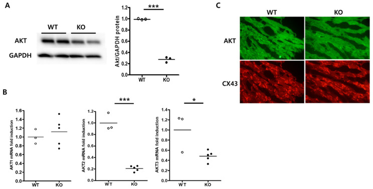
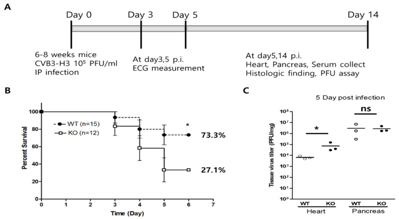
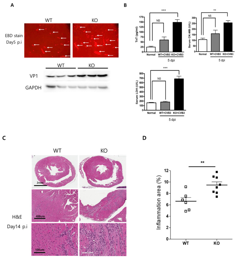
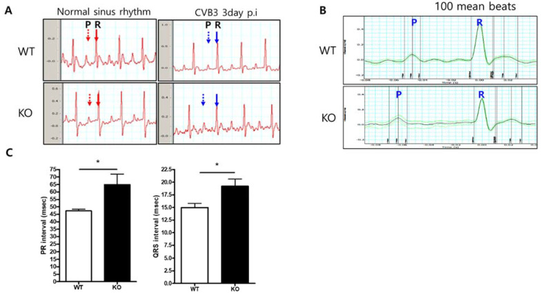
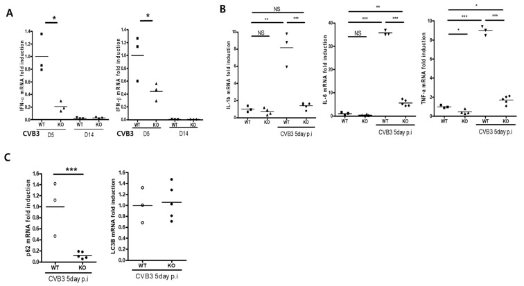
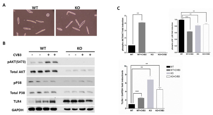
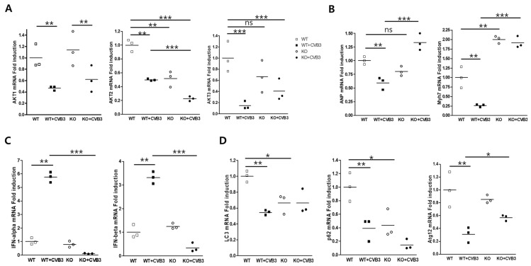
Protein kinase B2 (AKT2) is involved in various cardiomyocyte signaling processes, including those important for survival and metabolism. Coxsackievirus B3 (CVB3) is one of the most common pathogens that cause myocarditis in humans. The role of AKT2 in CVB3 infection is not yet well understood. We used a cardiac-specific AKT2 knockout (KO) mouse to determine the role of AKT2 in CVB3-mediated myocarditis. CVB3 was injected intraperitoneally into wild-type (WT) and KO mice. The mice's survival rate was recorded: survival in KO mice was significantly decreased compared with WT mice (WT vs. KO: 73.3 vs. 27.1%). Myocardial damage and inflammation were significantly increased in the hearts of KO mice compared with those of WT mice. Moreover, from surface ECG, AKT2 KO mice showed a prolonged atria and ventricle conduction time (PR interval, WT vs. KO: 47.27 ± 1.17 vs. 64.79 ± 7.17 ms). AKT2 deletion induced severe myocarditis and cardiac dysfunction due to CVB3 infection. According to real-time PCR, the mRNA level of IL-1, IL-6, and TNF-α decreased significantly in KO mice compared with WT mice on Days 5 after infection. In addition, innate immune response antiviral effectors, Type I interferon (interferon-α and β), and p62, were dramatically suppressed in the heart of KO mice. In particular, the adult cardiac myocytes isolated from the heart showed high induction of TLR4 protein in KO mice in comparison with WT. AKT2 deletion suppressed the activation of Type I interferon and p62 transcription in CVB3 infection. In cardiac myocytes, AKT2 is a key signaling molecule for the heart from damage through the activation of innate immunity during acute myocarditis.
Keywords: coxsackievirus B3; innate immunity; myocarditis; protein kinase B2; toll-like-receptor4.
Conflict of interest statement
The authors declare no conflict of interest.
Figures
Similar articles
-
Macrophage-Specific Coxsackievirus and Adenovirus Receptor Deletion Enhances Macrophage M1 Polarity in CVB3-Induced Myocarditis.Int J Mol Sci. 2023 Mar 10;24(6):5309. doi: 10.3390/ijms24065309. Int J Mol Sci. 2023. PMID: 36982385 Free PMC article.
-
Protease-activated receptor-2 regulates the innate immune response to viral infection in a coxsackievirus B3-induced myocarditis.J Am Coll Cardiol. 2013 Nov 5;62(19):1737-45. doi: 10.1016/j.jacc.2013.05.076. Epub 2013 Jul 17. J Am Coll Cardiol. 2013. PMID: 23871888 Free PMC article.
-
Adiponectin promotes coxsackievirus B3 myocarditis by suppression of acute anti-viral immune responses.Basic Res Cardiol. 2014 May;109(3):408. doi: 10.1007/s00395-014-0408-y. Epub 2014 Apr 2. Basic Res Cardiol. 2014. PMID: 24691762
-
Manipulating intestinal immunity and microflora: an alternative solution to viral myocarditis?Future Microbiol. 2012 Oct;7(10):1207-16. doi: 10.2217/fmb.12.96. Future Microbiol. 2012. PMID: 23030425 Review.
-
Immunopathological basis of virus-induced myocarditis.Clin Dev Immunol. 2004 Mar;11(1):1-5. doi: 10.1080/10446670410001670427. Clin Dev Immunol. 2004. PMID: 15154605 Free PMC article. Review.
Cited by
-
Macrophage-Specific Coxsackievirus and Adenovirus Receptor Deletion Enhances Macrophage M1 Polarity in CVB3-Induced Myocarditis.Int J Mol Sci. 2023 Mar 10;24(6):5309. doi: 10.3390/ijms24065309. Int J Mol Sci. 2023. PMID: 36982385 Free PMC article.
-
Circular RNA circ_0076631 promotes coxsackievirus B3 infection through modulating viral translation by sponging miR-214-3p.Front Microbiol. 2022 Sep 6;13:975223. doi: 10.3389/fmicb.2022.975223. eCollection 2022. Front Microbiol. 2022. PMID: 36147837 Free PMC article.
-
Functions of Circular RNA in Human Diseases and Illnesses.Noncoding RNA. 2023 Jul 4;9(4):38. doi: 10.3390/ncrna9040038. Noncoding RNA. 2023. PMID: 37489458 Free PMC article. Review.
-
Sex differences in the cardiac stress response following SARS-CoV-2 infection of ferrets.Am J Physiol Heart Circ Physiol. 2023 Nov 1;325(5):H1153-H1167. doi: 10.1152/ajpheart.00101.2023. Epub 2023 Sep 22. Am J Physiol Heart Circ Physiol. 2023. PMID: 37737732 Free PMC article.
References
-
- Baboonian C., Davies M.J., Booth J.C., McKenna W.J. Coxsackie B viruses and human heart disease. Curr. Top. Microbiol. Immunol. 1997;223:31–52. - PubMed
-
- Xiong D., Yajima T., Lim B.K., Stenbit A., Dublin A., Dalton N.D., Summers-Torres D., Molkentin J.D., Duplain H., Wessely R., et al. Inducible cardiac-restricted expression of enteroviral protease 2A is sufficient to induce dilated cardiomyopathy. Circulation. 2007;115:94–102. doi: 10.1161/CIRCULATIONAHA.106.631093. - DOI - PubMed
MeSH terms
Substances
Grants and funding
LinkOut - more resources
Full Text Sources
Molecular Biology Databases
Research Materials
Miscellaneous
