. 2022 Jan 28;23(3):1522.
doi: 10.3390/ijms23031522.

Induction of PLXNA4 Gene during Neural Differentiation in Human Umbilical-Cord-Derived Mesenchymal Stem Cells by Low-Intensity Sub-Sonic Vibration

Affiliations

Induction of PLXNA4 Gene during Neural Differentiation in Human Umbilical-Cord-Derived Mesenchymal Stem Cells by Low-Intensity Sub-Sonic Vibration

Hyunjin Cho et al. Int J Mol Sci. .

Abstract

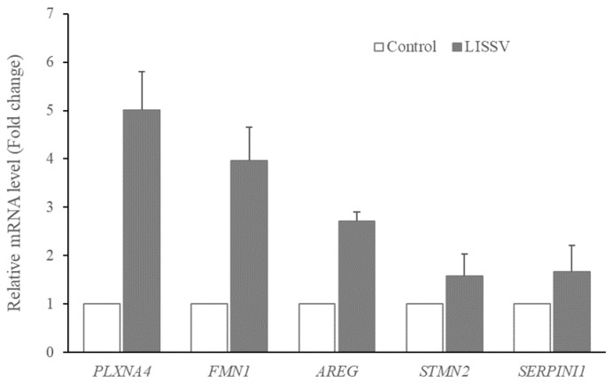
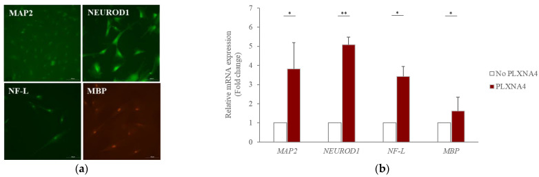
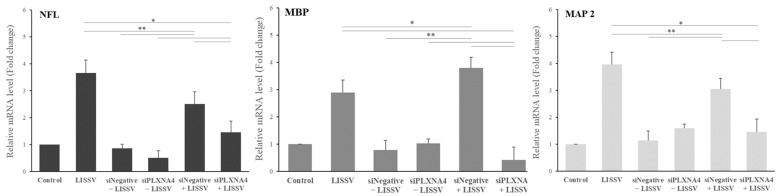
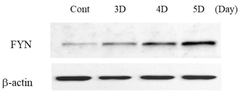
Human umbilical-cord-derived mesenchymal stem cells (hUC-MSC) are a type of mesenchymal stem cells and are more primitive than other MSCs. In this study, we identify novel genes and signal-activating proteins involved in the neural differentiation of hUC-MSCs induced by Low-Intensity Sub-Sonic Vibration (LISSV). RNA sequencing was used to find genes involved in the differentiation process by LISSV. The changes in hUC-MSCs caused by LISSV were confirmed by PLXNA4 overexpression and gene knockdown through small interfering RNA experiments. The six genes were increased among genes related to neurons and the nervous system. One of them, the PLXNA4 gene, is known to play a role as a guide for axons in the development of the nervous system. When the PLXNA4 recombinant protein was added, neuron-related genes were increased. In the PLXNA4 gene knockdown experiment, the expression of neuron-related genes was not changed by LISSV exposure. The PLXNA4 gene is activated by sema family ligands. The expression of SEMA3A was increased by LISSV, and its downstream signaling molecule, FYN, was also activated. We suggest that the PLXNA4 gene plays an important role in hUC-MSC neuronal differentiation through exposure to LISSV. The differentiation process depends on SEMA3A-PLXNA4-dependent FYN activation in hUC-MSCs.

Keywords: PLXNA4; SEMA3A; hUC-MSCs; low-intensity sub-sonic vibration; neural signaling.


Conflict of interest statement

The authors declare no conflict of interest.

Figures

Figure 1
Figure 1
Expression of genes in human umbilical-cord-derived mesenchymal stem cells after low-intensity sub-sonic vibration treatment using RNA sequencing. Cells were harvested 4 days after LISSV treatment. Fold change in expression of Plexin-A4 (PLXNA4), Formin 1 (FMN1), Amphiregulin (AREG), Stathmin 2 (STMN2), and Serpin Family I Member 1 (SERPINI1). Assayed using real-time polymerase chain reaction. Column heights correspond to mean values and error bars to standard deviations (n = 3).
Figure 2
Figure 2
Expression of each protein in human umbilical-cord-derived mesenchymal stem cells after low-intensity sub-sonic vibration treatment. Proteins detected were Serpin Family I Member 1 (SERPINI1), Plexin A4 (PLXNA4), Formin 1 (FMN1), and Amphiregulin (AREG). (a) Western blot image. (b) Intensities of each Western blot band were quantified by Image J. Each band was normalized using β-actin. Column heights correspond to mean values and error bars to standard deviations (n = 3). * p < 0.05, ** p < 0.01.
Figure 3
Figure 3
PLXNA4 gene expression after recombinant PLXNA4 protein treatment in human umbilical-cord-derived mesenchymal stem cells. Cells were harvested 4 days after recombinant PLXNA4 protein treatment. (a) Real-time PCR data of each protein. (b) MTT data show the inhibition of proliferation. Column heights correspond to mean values and error bars to standard deviations (n = 3). * p < 0.05, ** p < 0.01.
Figure 4
Figure 4
Morphological changes after recombinant PLXNA4 protein treatment for 5 days in human umbilical-cord-derived mesenchymal stem cells. (a1,a2): Untreated control cells. (b1,b2): 1.5 μg of protein-treated cells. (c1,c2): 2.0 μg of protein-treated cells. Original magnification 40×.
Figure 5
Figure 5
Expression of neuron-related proteins and genes in human umbilical-cord-derived mesenchymal stem cells after recombinant PLXNA4 protein treatment. Cells were harvested 5 days after 2 μg/mL recombinant PLXNA4 protein treatment. (a) Fluorescence images of each protein. Original magnification 200×. (b) Fold expression of each gene using real-time polymerase chain reaction analysis. MAP2: Microtubule-associated protein 2. NEUROD1: Neuronal Differentiation 1, NF-L: Neurofilament-L, MBP: Myelin basic protein, Column heights correspond to mean values and error bars to standard deviations (n = 3). * p < 0.05, ** p < 0.01.
Figure 6
Figure 6
Expression of calcium channels in human umbilical-cord-derived mesenchymal stem cells after recombinant PLXNA4 protein treatment. Cells were harvested 5 days after 2 μg/mL recombinant PLXNA4 protein treatment. Fold expression of each gene using real-time polymerase chain reaction analysis. Cav2.1: Voltage-gated P/Q type calcium channel. Cav2.2: Voltage-gated N-type calcium channel, Column heights correspond to mean values and error bars to standard deviations (n = 3). ** p < 0.01.
Figure 7
Figure 7
MTT data for siRNA transfection in human umbilical-cord-derived mesenchymal stem cells. Cells were harvested 2 days after siRNA duplex transfection. Column heights correspond to mean values and error bars to standard deviations (n = 3). * p < 0.05.
Figure 8
Figure 8
PLXNA4 gene silencing using small interfering RNA transfection methods in human umbilical-cord-derived mesenchymal stem cells. Cells were harvested 4 days after low-intensity sub-sonic vibration treatment. (a) Morphology of each sample: A. Control; B. low-intensity sub-sonic vibration (LISSV); C. siNegative duplex 90 nmol without (−) LISSV; D. siPLXNA4 duplex 90 nmol without (−) LISSV; E. siNegative duplex with (+) LISSV; F. siPLXNA4 duplex with (+) LISSV. Original magnification 40×. (b) Fold change in the expression PLXNA4 determined using real-time polymerase chain reaction analysis. Column heights correspond to mean values and error bars to standard deviations (n = 3). * p < 0.05, ** p < 0.01.
Figure 9
Figure 9
Expression of neuron-associated genes in human umbilical-cord-derived mesenchymal stem cells after siPLXNA4 duplex transfection using real-time polymerase chain reaction analysis. Cells were harvested 4 days after low-intensity sub-sonic vibration treatment. NF-L: Neurofilament-L, MBP: Myelin basic protein, MAP2: Microtubule-associated protein 2. Column heights correspond to mean values and error bars to standard deviations (n = 3). * p < 0.05, ** p < 0.01.
Figure 10
Figure 10
Expression of neural differentiation-specific genes in human umbilical-cord-derived mesenchymal stem cells after low-intensity sub-sonic vibration versus recombinant PLXNA4 protein. Cells were harvested 4 days after low-intensity sub-sonic vibration treatment and 5 days after recombinant PLXNA4 protein treatment. The fold change in the expression of each gene was analyzed using a real-time polymerase chain reaction. MAP2: Microtubule-associated protein 2. NEUROD1: Neuronal Differentiation 1, GFAP: Glial Fibrillary Acidic Protein, MBP: Myelin basic protein. Column heights correspond to mean values and error bars to standard deviations (n = 3). * p < 0.05, ** p < 0.01.
Figure 11
Figure 11
The changes of specific markers in human umbilical-cord-derived mesenchymal stem cells by low-intensity sub-sonic vibration and recombinant PLXNA4 protein treatment using FACS analysis. Cells were harvested 3 days after each treatment. PE-conjugated anti-CD73 and PE-conjugated anti-CD105 were used. (A,D) Control. (B,E) LISSV treatment. (C,F) 2 μg/mL recombinant PLXNA4 protein treatment.
Figure 12
Figure 12
Expression of PLXNA4-dependent semaphorin signaling molecules in human umbilical-cord-derived mesenchymal stem cells by low-intensity sub-sonic vibration using real-time polymerase chain reaction analysis. Cells were harvested 4 days after low-intensity sub-sonic vibration treatment. SEMA3A: Semaphorin 3A, SEMA6A: Semaphorin 6A. Column heights correspond to mean values and error bars to standard deviations (n = 3).
Figure 13
Figure 13
Expression of PLXNA4-dependent semaphorin signaling protein in human umbilical-cord-derived mesenchymal stem cells by low-intensity sub-sonic vibration. FYN: Src Family of nonreceptor tyrosine kinase p59.
Figure 14
Figure 14
Expression of presynaptic vesicle protein-associated genes in human umbilical-cord-derived mesenchymal stem cells after low-intensity sub-sonic vibration versus recombinant PLXNA4 protein. Cells were harvested 4 days after low-intensity sub-sonic vibration treatment and 5 days after recombinant PLXNA4 protein treatment. The fold change in the expression of each gene was analyzed using real-time polymerase chain reaction. SYN1: Synapsin 1, GAP43: Growth-Associated Protein 43, Column heights correspond to mean values and error bars to standard deviations (n = 3). * p < 0.05, ** p < 0.01.

Similar articles

Cited by

References

    1. Huang Y., Jia X., Bai K., Gong X., Fan Y. Effect of Fluid Shear Stress on Cardiomyogenic Differentiation of Rat Bone Marrow Mesenchymal Stem Cells. Arch. Med. Res. 2010;41:497–505. doi: 10.1016/j.arcmed.2010.10.002. - DOI - PubMed
    1. Maul T.M., Chew D.W., Nieponice A., Vorp D.A. Mechanical stimuli differentially control stem cell behavior: Morphology, proliferation, and differentiation. Biomech. Model. Mechanobiol. 2011;10:939–953. doi: 10.1007/s10237-010-0285-8. - DOI - PMC - PubMed
    1. Kim D.H., Heo S.J., Kim S.H., Shin J.W., Park S.H., Shin J.W. Shear stress magnitude is critical in regulating the differentiation of mesenchymal stem cells even with endothelial growth medium. Biotechnol. Lett. 2011;33:2351–2359. doi: 10.1007/s10529-011-0706-5. - DOI - PubMed
    1. Dan P., Velot É., Decot V., Menu P. The role of mechanical stimuli in the vascular differentiation of mesenchymal stem cells. J. Cell Sci. 2015;128:2415–2422. doi: 10.1242/jcs.167783. - DOI - PubMed
    1. Fahy N., Alini M., Stoddart M.J. Mechanical stimulation of mesenchymal stem cells: Implications for cartilage tissue engineering. J. Orthop. Res. 2018;36:52–63. doi: 10.1002/jor.23670. - DOI - PubMed

Substances

LinkOut - more resources