Review
. 2022 Jan 29;23(3):1580.
doi: 10.3390/ijms23031580.

Polymodal Control of TMEM16x Channels and Scramblases

Affiliations
Review

Polymodal Control of TMEM16x Channels and Scramblases

Emilio Agostinelli et al. Int J Mol Sci. .

Abstract

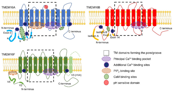
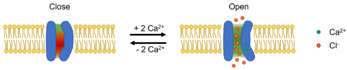
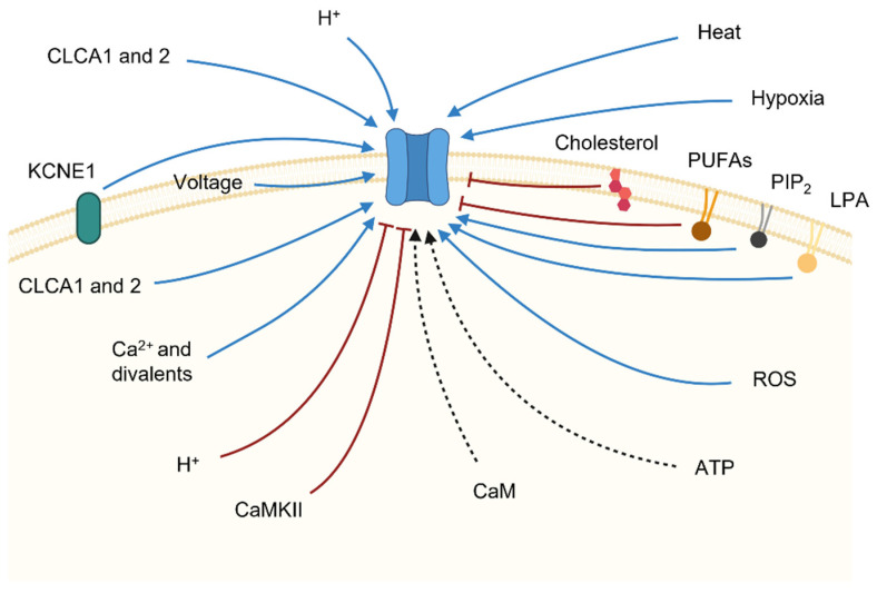
The TMEM16A/anoctamin-1 calcium-activated chloride channel (CaCC) contributes to a range of vital functions, such as the control of vascular tone and epithelial ion transport. The channel is a founding member of a family of 10 proteins (TMEM16x) with varied functions; some members (i.e., TMEM16A and TMEM16B) serve as CaCCs, while others are lipid scramblases, combine channel and scramblase function, or perform additional cellular roles. TMEM16x proteins are typically activated by agonist-induced Ca2+ release evoked by Gq-protein-coupled receptor (GqPCR) activation; thus, TMEM16x proteins link Ca2+-signalling with cell electrical activity and/or lipid transport. Recent studies demonstrate that a range of other cellular factors-including plasmalemmal lipids, pH, hypoxia, ATP and auxiliary proteins-also control the activity of the TMEM16A channel and its paralogues, suggesting that the TMEM16x proteins are effectively polymodal sensors of cellular homeostasis. Here, we review the molecular pathophysiology, structural biology, and mechanisms of regulation of TMEM16x proteins by multiple cellular factors.

Keywords: Ca2+ signalling; Ca2+-activated Cl− channels; SARS-CoV-2; TMEM16x; anoctamin; gating; lipids; scramblases.


Conflict of interest statement

The authors declare no conflict of interest.

Figures

Figure 1
Figure 1
Topological diagram of TMEM16x proteins, showing the position of the alternative spliced exons and a range of functional domains. The boxes (broken lines) highlight the TMs involved in the formation of the ion/lipid pathways in TMEM16x. The approximate position of each splicing variant is labelled using the nomenclature described in the main text. The diagrams also show the approximate position of a range of functional domains, including (1) the principal Ca2+-binding pocket, which encompasses conserved acidic residues located in TM6-8; (2) additional regulatory Ca2+-binding sites; (3) putative calmodulin (CaM)-binding sites; (4) putative phosphatidylinositol 4,5-bisphosphate (PIP2)-binding sites; and (5) the pH-sensitive domain on the extracellular loop between TM5 and TM6 of TMEM16A.
Figure 2
Figure 2
Structural alignment of the experimentally determined Ca2+-bound structure of TMEM16A channel, and a model of the TMEM16A channel in the open state. (A) Left: Lateral view of the dimeric Ca2+-bound TMEM16A cryo-EM structure (PDB ID: 5OYB, blue). Right: TMEM16A open-state model (from [122], grey). The Ca2+ ions are shown in green. (B) Left: Single monomer of the Ca2+-bound TMEM16A cryo-EM structure (5OYB, blue), aligned with the open-state model (grey) [122]. The Ca2+ ions are shown in green. Right: Magnification of the gate-forming pore residues (V539 and I636, from [122]), in the Ca2+-bound TMEM16A cryo-EM structure (5OYB, blue), and of the TMEM16A open-state model (grey) [122], as indicated.
Figure 3
Figure 3
TMEM16A close and open conformation diagram. Diagrammatic representation of a single TMEM16A channel pore and related gating mechanisms. Ca2+ binding induces opening of the steric gate in TM6 and attenuation of the electrostatic gate. The movement of the TM6 helix during gating is represented as a tilt of the inner portion of the pore and opening of the outer pore. The green and red backgrounds depict positive and negative electrostatic potentials in the pore, respectively.
Figure 4
Figure 4
Summary of the principal factors that control TMEM16x function. Schematic representation of various physiological modulators of TMEM16x activity. A TMEM16x protein in the plasma membrane of a generic cell is shown in blue. Factors that control TMEM16x activity are connected to the protein with red or blue lines to denote inhibitory or stimulatory influences, respectively. The black dashed arrows indicate the modulations for which an underlying mechanism has not yet been fully defined and/or conflicting evidence has been proposed.

Similar articles

Cited by

References

    1. Schroeder B.C., Cheng T., Jan Y.N., Jan L.Y. Expression Cloning of TMEM16A as a Calcium-Activated Chloride Channel Subunit. Cell. 2008;134:1019–1029. doi: 10.1016/j.cell.2008.09.003. - DOI - PMC - PubMed
    1. Bushell S.R., Pike A.C.W., Falzone M.E., Rorsman N.J.G., Ta C.M., Corey R.A., Newport T.D., Christianson J.C., Scofano L.F., Shintre C.A., et al. The Structural Basis of Lipid Scrambling and Inactivation in the Endoplasmic Reticulum Scramblase TMEM16K. Nat. Commun. 2019;10:3956. doi: 10.1038/s41467-019-11753-1. - DOI - PMC - PubMed
    1. Di Zanni E., Gradogna A., Scholz-Starke J., Boccaccio A. Gain of Function of TMEM16E/ANO5 Scrambling Activity Caused by a Mutation Associated with Gnathodiaphyseal dysplasia. Cell. Mol. Life Sci. 2018;75:1657–1670. doi: 10.1007/s00018-017-2704-9. - DOI - PMC - PubMed
    1. Guo J., Wang D., Dong Y., Gao X., Tong H., Liu W., Zhang L., Sun M. ANO7: Insights into Topology, Function, and Potential Applications as a Biomarker and Immunotherapy Target. Tissue Cell. 2021;72:101546. doi: 10.1016/j.tice.2021.101546. - DOI - PubMed
    1. Kim H., Kim H., Lee J., Lee B., Kim H.-R., Jung J., Lee M.-O., Oh U. Anoctamin 9/TMEM16J Is a Cation Channel Activated by CAMP/PKA Signal. Cell Calcium. 2018;71:75–85. doi: 10.1016/j.ceca.2017.12.003. - DOI - PubMed

LinkOut - more resources