. 2022 Jan 29;23(3):1599.
doi: 10.3390/ijms23031599.

The Enhanced Cytotoxic Effects in B-Cell Leukemia and Lymphoma Following Activation of Prostaglandin EP4 Receptor and Targeting of CD20 Antigen by Monoclonal Antibodies

Affiliations

The Enhanced Cytotoxic Effects in B-Cell Leukemia and Lymphoma Following Activation of Prostaglandin EP4 Receptor and Targeting of CD20 Antigen by Monoclonal Antibodies

Tijana Markovič et al. Int J Mol Sci. .

Abstract

Anti-CD20 monoclonal antibodies (MAbs) have revolutionized the treatment of B-cell leukemia and lymphoma. However, many patients do not respond to such treatment due to either deficiency of the complementary immune response or resistance to apoptosis. Other currently available treatments are often inadequate or induce major side effects. Therefore, there is a constant need for improved therapies. The prostaglandin E2 receptor 4 (EP4) receptor has been identified as a promising therapeutic target for hematologic B-cell malignancies. Herein, we report that EP4 receptor agonists PgE1-OH and L-902688 have exhibited enhanced cytotoxicity when applied together with anti-CD20 MAbs rituximab, ofatumumab and obinutuzumab in vitro in Burkitt lymphoma cells Ramos, as well as in p53-deficient chronic lymphocytic leukemia (CLL) cells MEC-1. Moreover, the enhanced cytotoxic effects of EP4 receptor agonists and MAbs targeting CD20 have been identified ex vivo on primary lymphocytes B obtained from patients diagnosed with CLL. Incubation of cells with PgE1-OH and L-902688 preserved the expression of CD20 molecules, further confirming the anti-leukemic potential of EP4 receptor agonists in combination with anti-CD20 MAbs. Additionally, we demonstrated that the EP4 receptor agonist PgE-1-OH induced apoptosis and inhibited proliferation via the EP4 receptor triggering in CLL. This work has revealed very important findings leading towards the elucidation of the anticancer potential of PgE1-OH and L-902688, either alone or in combination with MAbs. This may contribute to the development of potential therapeutic alternatives for patients with B-cell malignancies.

Keywords: B-cell leukemia and lymphoma; chronic lymphocytic leukemia; monoclonal antibodies; prostaglandin EP4 receptor; selective EP4 receptor agonist; synergistic effects.


Conflict of interest statement

The authors declare no conflict of interest.

Figures

Figure 1
Figure 1
Anti-CD20 MAbs rituximab and ofatumumab induce CDC while obinutuzumab is less effective. The effect of active human serum on MAbs induced CDC was evaluated in Ramos (A) and MEC-1 (B) cells. The cells were incubated with a therapeutic concentration of 10 µg/mL of rituximab, ofatumumab or obinutuzumab in the presence of 1%, 5% or 20% active human serum and their metabolic activities were determined after 24 h. MAbs-induced CDC was assessed in Ramos (C) and MEC-1 (D) cells. The cells were incubated in the presence of 20% active human serum, heat deactivated human serum or 20% active human serum and eculizumab. Metabolic activity was determined after 24 h. Red dotted line represents the relative metabolic activity of untreated control cells. Data are presented as ratios relative to untreated control cells (mean ± SD of three independent experiments performed in triplicate). ANOVA, with post hoc analysis using Tukey’s multiple comparison, * p ˂ 0.05, *** p ˂ 0.001, **** p ˂ 0.0001.
Figure 2
Figure 2
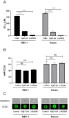
The cytotoxic effects of PgE1-OH and L-902688 are transduced via EP4 receptor. (A) MEC-1 and Ramos cells were exposed to increasing concentrations of PGE2, PgE1-OH, L-902688 (i.e., 1, 5, 10, 25, 50, 100, 150 μM). After 24 h, the metabolic activity of cells was determined and the IC50 values were calculated. Data are presented as ratios relative to untreated control cells (mean ± SD of three independent experiments performed in triplicate). Student’s t-test, ****, p < 0.0001, ns, not significant. (B) CD20 expression is preserved after EP4 receptor triggering. CD20 expression was determined in MEC-1 and Ramos cells. Cells were treated with non-cytotoxic concentrations of PgE1-OH (5 µM) and L-902688 (1 or 2.5 µM) for 24 h or left untreated (control cells). Median fluorescence intensity (MFI) of CD20 antigen was evaluated by flow cytometry (mean ± SD of three independent experiments). Student’s t-test, ns denotes p ≥ 0.05. (C) The expression of CD20 was determined by imaging flow cytometer and the images of representative cells are presented. Scale bar indicates 7 µm.
Figure 3
Figure 3
PgE1-OH induces apoptosis in CLL cells MEC-1. (A) Dose-dependent increases in the percentage of cells undergoing early and late apoptosis as compared to the untreated control. Cells were stained with Annexin V and SytoxBlue. Cells in the lower left quadrant are Annexin V-negative/SytoxBlue-negative (viable), cells in the lower right quadrant are Annexin V-positive/SytoxBlue-negative (pro-apoptotic), whereas cells in the upper right quadrant are Annexin V-positive/SytoxBlue-positive (late apoptotic). (B) Percentage of cells undergoing early apoptosis induced by PgE1-OH (mean ± SEM of two independent experiments). One-way ANOVA, with post hoc analysis using Tukey’s multiple comparison, *, p ˂ 0.05, ** p ˂ 0.01. (C) Anti-proliferative activity of PgE1-OH on MEC-1 cells. MEC-1 cells were stained with CFSE dye and incubated with 10 μM PgE1-OH or the vehicle treated control (0.1% DMSO) for 0, 24, 48 or 72 h. The proliferation of PgE1-OH-treated and untreated (0.1% DMSO) cells was analyzed by flow cytometry. Peaks 0, 1, 2, 3 represent fluorescence of CFSE stained cells after incubation periods of 0, 24, 48 or 72 h, respectively.
Figure 4
Figure 4
EP4 receptor agonists enhance the cytotoxicity of anti-CD20 MAbs against Burkitt lymphoma cell line Ramos. Cells were incubated with 10 μM PgE1-OH (A,C,E), 5 μM L-902688 (B,D,F) and 1 μg/mL or 10 μg/mL rituximab (A,B), ofatumumab (C,D) or obinutuzumab (E,F) in the presence of 1% human serum. Metabolic activity was determined after 48 h. Red dotted line represents the relative metabolic activity of untreated control cells. Data are presented as ratios relative to untreated control cells (mean ± SD of three independent experiments performed in triplicate). ANOVA, with post hoc analysis using Tukey’s multiple comparison, *, p ˂ 0.05, ** p ˂ 0.01, *** p ˂ 0.001, **** p ˂ 0.0001.
Figure 5
Figure 5
EP4 receptor agonists enhance cytotoxicity of anti-CD20 MAbs against the CLL cell line MEC-1. Cells were incubated with 10 μM PgE1-OH (A,C,E), 5 μM L-902688 (B,D,F) and 1 or 10 μg/mL rituximab (A,B), ofatumumab (C,D) or obinutuzumab (E,F) in the presence of one percent human serum. Metabolic activity was determined after 48 h. Red dotted line represents the relative metabolic activity of untreated control cells. Data are presented as ratios relative to untreated control cells (mean ± SD of three independent experiments performed in triplicate). ANOVA, with post hoc analysis using Tukey´s multiple comparison, *, p ˂ 0.05, *** p ˂ 0.001.
Figure 6
Figure 6
EP4 receptor agonist-induced cytotoxicity in primary CLL cells is augmented by anti-CD 20 MAbs rituximab and ofatumumab. (A) The degree of MAbs-induced CDC was evaluated in primary CLL cells (n = 7). The impact of concentration of autologous human serum on MAbs-induced CDC was determined by cultivating the cells at therapeutic concentrations of 10 μg/mL of rituximab, ofatumumab and obinutuzumab in the presence of 1%, 5% and 20% autologous serum. Metabolic activity was determined after 48 h. Data are presented as ratios relative to untreated control cells (mean ± SD, from seven samples of unrelated CLL donors, performed in triplicate). (B) CD20 expression was determined in primary CLL cells (n = 9). Cells were treated with non-cytotoxic concentrations of (10 µM) and L-902688 (2.5 µM) for 24 h or left untreated (control cells). Percentage of CD20 antigen positive cells was evaluated by flow cytometry (mean ± SEM, from nine CLL samples). Synergistic effects of PgE1-OH and (C) rituximab, (D) ofatumumab in primary CLL cells. CLL cells were incubated with PgE1-OH (10 μM) and MAbs (1 μg/mL, 10 μg/mL) in the presence of one percent autologous human serum. Metabolic activity was determined after 48 h. The experiments were performed in triplicate. Red dotted line represents the relative metabolic activity of untreated control cells. Data are presented as ratios relative to untreated control cells. Each sign denotes a sample of a representative CLL donor (n = 4).

References

    1. Torre L.A., Bray F., Siegel R.L., Ferlay J., Lortet-Tieulent J., Jemal A. Global Cancer Statistics, 2012. CA Cancer J. Clin. 2015;65:87–108. doi: 10.3322/caac.21262. - DOI - PubMed
    1. Salles G., Barrett M., Foà R., Maurer J., O’Brien S., Valente N., Wenger M., Maloney D.G. Rituximab in B-Cell Hematologic Malignancies: A Review of 20 Years of Clinical Experience. Adv. Ther. 2017;34:2232–2273. doi: 10.1007/s12325-017-0612-x. - DOI - PMC - PubMed
    1. Middleton O., Cosimo E., Dobbin E., McCaig A.M., Clarke C.L., Brant A.M., Leach M., Michie A., Wheadon H. Complement deficiencies limit CD20 monoclonal antibody treatment efficacy in CLL. Leukemia. 2015;29:107–114. doi: 10.1038/leu.2014.146. - DOI - PubMed
    1. Byrd J.C., Stilgenbauer S., Flinn I.W. Chronic Lymphocytic Leukemia. Hematology. 2004;2004:163–183. doi: 10.1182/asheducation-2004.1.163. - DOI - PubMed
    1. Glennie M.J., French R.R., Cragg M.S., Taylor R.P. Mechanisms of killing by anti-CD20 monoclonal antibodies. Mol. Immunol. 2007;44:3823–3837. doi: 10.1016/j.molimm.2007.06.151. - DOI - PubMed

MeSH terms