Comparative Study
. 2022 Jan 30;23(3):1613.
doi: 10.3390/ijms23031613.

New Evidence of the Importance of Weak Interactions in the Formation of PML-Bodies

Affiliations
Comparative Study

New Evidence of the Importance of Weak Interactions in the Formation of PML-Bodies

Alexander V Fonin et al. Int J Mol Sci. .

Abstract

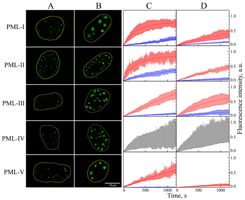
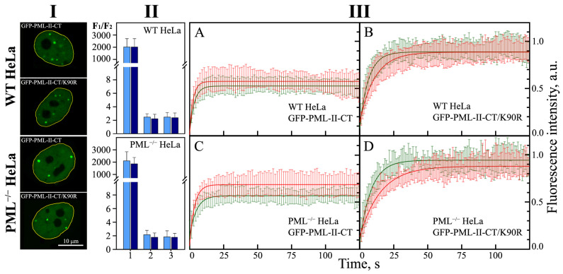
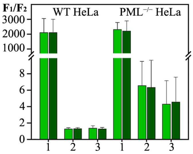
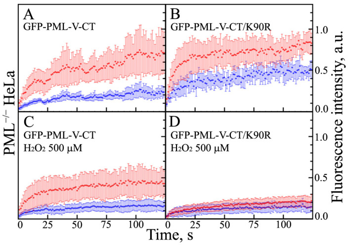
In this work, we performed a comparative study of the formation of PML bodies by full-length PML isoforms and their C-terminal domains in the presence and absence of endogenous PML. Based on the analysis of the distribution of intrinsic disorder predisposition in the amino acid sequences of PML isoforms, regions starting from the amino acid residue 395 (i.e., sequences encoded by exons 4-6) were assigned as the C-terminal domains of these proteins. We demonstrate that each of the full-sized nuclear isoforms of PML is capable of forming nuclear liquid-droplet compartments in the absence of other PML isoforms. These droplets possess dynamic characteristics of the exchange with the nucleoplasm close to those observed in the wild-type cells. Only the C-terminal domains of the PML-II and PML-V isoforms are able to be included in the composition of the endogenous PML bodies, while being partially distributed in the nucleoplasm. The bodies formed by the C-terminal domain of the PML-II isoform are dynamic liquid droplet compartments, regardless of the presence or absence of endogenous PML. The C-terminal domain of PML-V forms dynamic liquid droplet compartments in the knockout cells (PML-/-), but when the C-terminus of the PML-V isoform is inserted into the existing endogenous PML bodies, the molecules of this protein cease to exchange with the nucleoplasm. It was demonstrated that the K490R substitution, which disrupts the PML sumoylation, promotes diffuse distribution of the C-terminal domains of PML-II and PML-V isoforms in endogenous PML knockout HeLa cells, but not in the wild-type cells. These data indicate the ability of the C-terminal domains of the PML-II and PML-V isoforms to form dynamic liquid droplet-like compartments, regardless of the ordered N-terminal RBCC motifs of the PML. This indicates a significant role of the non-specific interactions between the mostly disordered C-terminal domains of PML isoforms for the initiation of liquid-liquid phase separation (LLPS) leading to the formation of PML bodies.

Keywords: PML-bodies; acute hydrogen peroxide-induced oxidative stress; fluorescence recovery after photobleaching (FRAP); liquid–liquid phase separation (LLPS); membrane-less organelles (MLOs); promyelocytic leukemia protein (PML) isoforms.


Conflict of interest statement

The authors declare no conflict of interest.

Figures

Figure 1
Figure 1
Localization, shape, and dynamic properties of PML bodies visualized by various exogenous EGFP-PML isoforms in the absence of all endogenous forms of PML protein in PML−/− HeLa cells. Nuclear localization of normal, small (area < 1.5 μm2), and large (area > 1.75 μm2) PML-bodies with exogenous PML isoforms I–V are represented in Panel (A,B), respectively. Panel (C,D): Curves of EGFP fluorescence recovery after photobleaching of nuclear PML isoforms in PML-bodies in studied cells in absence of stress and in acute oxidative stress conditions, respectively. The curves of the photoreduction of PML isoforms in the composition of “small” PML bodies are shown in red, the curves of the photoreduction of PML isoforms in the composition of “large” PML bodies are shown in blue, and the curves of the photoreduction of PML-IV isoforms are shown in gray. Solid curves represent the fit of FRAP data in the framework of the mono-exponential approximation. The standard deviations of the data are shown by the error bars of the corresponding color.
Figure 2
Figure 2
Colocalization of endogenous PML-bodies and C-terminal domains of PML isoforms in the HeLa cells using confocal fluorescent microscopy. Nucleus are colored by DAPI (column (A)). C-terminal domains of PML isoforms are visualized by expression of chimeric proteins EGFP fused with C-terminal domain of appropriate PML isoform (column (B)). The endogenous PML-bodies are visualized by cells staining with Anti-PML Alexa Fluor 647 antibodies (column (C)). Merging of images in columns (AC) (column (D)).
Figure 3
Figure 3
Localization, shape, and dynamic properties of the condensates formed by the C-terminal domain of PML-II isoform (PML-II-CT) and its mutant form with the K490R amino acid replacement (PML-II-CT/K490R) in wild-type HeLa cells (WT HeLa) and PML knockout HeLa cells (PML−/− HeLa). Panel (I): Nuclear localization of condensates formed by PML-II-CT and PML-II-CT/K490R in the WTHeLa and PML−/− HeLa. Panel (II): Relative contents of the isoform PML-II, PML-II-CT and PML-II-CT/K490R in bodies formed by these proteins and the nucleoplasm in the WT HeLa (upper panel), and the PML−/− HeLa (lower panel) are shown by bars 1, 2 and 3, respectively. Light blue bars correspond to the normal conditions and that dark blue bars to H2O2-treated cells. The concentration of exogenously expressed forms of PML was estimated by the ratio of the average fluorescence intensity observed in the bodies (F1) to the average fluorescence intensity in a region of the same size in the nucleoplasm (F2). Bars and error bars represent means and s.d. (n = 3). Panel (III): Curves of EGFP fluorescence recovery after photobleaching of PML-II-CT (panels (A,C)) and PML-II-CT/K490R (panels (B,D)) in the WT HeLa (panels (A,B)) and the PML−/− HeLa cells (panels (C,D)). The curves of the photoreduction of the target proteins at the normal conditions are shown in red, and the stress conditions (H2O2) are shown in green. Solid curves represent the fit of the FRAP data in the framework of the mono-exponential approximation. The standard deviations of the data are shown by the error bars of the corresponding color.
Figure 4
Figure 4
Relative contents of the isoform PML-V, PML-V-CT and PML-V-CT/K490R in bodies formed by these proteins and nucleoplasm in the WT HeLa (on the left), and in the PML−/− HeLa (on the right) are presented by bars 1, 2, and 3, respectively. Light green bars correspond to the normal conditions and that dark green bar correspond to the H2O2-treated cells. The concentration of exogenously expressed forms of PML was estimated by the ratio of the average fluorescence intensity observed in the bodies to the average fluorescence intensity in a region of the same size in the nucleoplasm. Bars and error bars represent means and s.d. (n = 3).
Figure 5
Figure 5
Dynamic properties of the condensates formed by the C-terminal domain of PML-V isoform and its mutant form with the K490R amino acid replacement in PML−/− HeLa cells. EGFP fluorescence recovery after photobleaching of C-terminal domain of PML-V isoform. Panels (A,C) present WT C-terminal domain of PMLV isoform and Panels (B,D) presents its mutant form with amino acid replacement K490R in normal (Panels (A,B)) and stressed (Panels (C,D)) conditions. The curves of the photoreduction of PML isoforms in the composition of “small” and “large” PML bodies are shown in red and blue. Solid curves represent the fit of the FRAP data in the framework of the mono-exponential approximation. The standard deviations of the data are shown by error bars of the corresponding color.

References

    1. Banani S.F., Lee H.O., Hyman A.A., Rosen M.K. Biomolecular condensates: Organizers of cellular biochemistry. Nat. Rev. Mol. Cell Biol. 2017;18:285–298. doi: 10.1038/nrm.2017.7. - DOI - PMC - PubMed
    1. Banani S.F., Rice A.M., Peeples W.B., Lin Y., Jain S., Parker R., Rosen M.K. Compositional Control of Phase-Separated Cellular Bodies. Cell. 2016;166:651–663. doi: 10.1016/j.cell.2016.06.010. - DOI - PMC - PubMed
    1. Bergeron-Sandoval L.P., Safaee N., Michnick S.W. Mechanisms and Consequences of Macromolecular Phase Separation. Cell. 2016;165:1067–1079. doi: 10.1016/j.cell.2016.05.026. - DOI - PubMed
    1. Berry J., Weber S.C., Vaidya N., Haataja M., Brangwynne C.P. RNA transcription modulates phase transition-driven nuclear body assembly. Proc. Natl. Acad. Sci. USA. 2015;112:E5237–E5245. doi: 10.1073/pnas.1509317112. - DOI - PMC - PubMed
    1. Boeynaems S., Alberti S., Fawzi N.L., Mittag T., Polymenidou M., Rousseau F., Schymkowitz J., Shorter J., Wolozin B., Van Den Bosch L., et al. Protein Phase Separation: A New Phase in Cell Biology. Trends Cell Biol. 2018;28:420–435. doi: 10.1016/j.tcb.2018.02.004. - DOI - PMC - PubMed

Publication types

MeSH terms

LinkOut - more resources