. 2022 Jan 31;23(3):1648.
doi: 10.3390/ijms23031648.

The Roles of Two CNG Channels in the Regulation of Ascidian Sperm Chemotaxis

Affiliations

The Roles of Two CNG Channels in the Regulation of Ascidian Sperm Chemotaxis

Kogiku Shiba et al. Int J Mol Sci. .

Abstract

Spermatozoa sense and respond to their environmental signals to ensure fertilization success. Reception and transduction of signals are reflected rapidly in sperm flagellar waveforms and swimming behavior. In the ascidian Ciona intestinalis (type A; also called C. robusta), an egg-derived sulfated steroid called SAAF (sperm activating and attracting factor), induces both sperm motility activation and chemotaxis. Two types of CNG (cyclic nucleotide-gated) channels, Ci-tetra KCNG (tetrameric, cyclic nucleotide-gated, K+-selective) and Ci-HCN (hyperpolarization-activated and cyclic nucleotide-gated), are highly expressed in Ciona testis from the comprehensive gene expression analysis. To elucidate the sperm signaling pathway to regulate flagellar motility, we focus on the role of CNG channels. In this study, the immunochemical analysis revealed that both CNG channels are expressed in Ciona sperm and localized to sperm flagella. Sperm motility analysis and Ca2+ imaging during chemotaxis showed that CNG channel inhibition affected the changes in flagellar waveforms and Ca2+ efflux needed for the chemotactic turn. These results suggest that CNG channels in Ciona sperm play a vital role in regulating sperm motility and intracellular Ca2+ regulation during chemotaxis.

Keywords: cAMP; cGMP; calcium; fertilization; sperm chemotaxis.


Conflict of interest statement

The authors declare no conflict of interest.

Figures
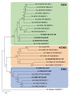
Figure 1
Figure 1
Phylogenetic analysis of cyclic nucleotide-gated channels from Ciona, sea urchins and other animals. The following ion channel sequences were used for the alignment: the HCN channel from Ciona (Ci-HCN1, 2, 3), sea urchin (Sp-HCN1, 2), human (Hs-HCN1, 2, 3, 4), zebrafish (Dr-HCN1, 2, 3), fruit fry (Dm-HCN), spiny lobster (Pa-HCN); the CNG channel from Ciona (Ci-CNG, 3, 4), sea urchin (Sp-CNG2), human (Hs-CNG1, 2, 3); CNGK channels from Ciona (Ci-tetraKCNG), sea urchin (Sp-tetraKCNG), acorn worm (Sk-CNGK), amphioxus (Bf-CNGK), starlet sea anemone (NvCNGK), zebrafish (Dr-CNGK) and rainbow trout (OmCNGK). The Drosophila shaker channel (Ds-Shaker) was used as an outgroup. The value on each branch represents the number of times that a node was supported in 100 bootstrap pseudoreplications.
Figure 2
Figure 2
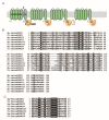
(A) Schematic representation of the tetraKCNG channel. * The asterisk shows the region used for the antigen to raise a polyclonal antibody. (B) Amino acid sequence alignment of the cyclic nucleotide-binding domain (CNBD) from the Sp-tetraKCNG channel and the Ci-tetraKCNG channel. The tetraKCNG channel identified in Ciona is very similar to the tetraKCNG channel in sea urchin. (C) Amino acid sequence alignment of the pore region.
Figure 3
Figure 3
(A) Schematic representation of the HCN channel. * The asterisk shows the region used for antigen to raise a polyclonal antibody. (B) Amino acid sequence alignment of the transmembrane segments S1–6 and the pore region from the Sp-HCN1, 2, Ci-HCN1 and 2. (C) Amino acid sequence alignment of the cyclic nucleotide-binding domain (CNBD) from the Sp-HCN1, 2, Ci-HCN1 and 2.
Figure 4
Figure 4
Tissue expression patterns of the CNG genes in Ciona tissues. RT-PCR analysis of mRNA from several adult Ciona tissues shows that the expression of KCNG, HCN1 and 2 is testis-specific. β-Actin was used as an internal control. E, endostyle; G, gill; T, testis; O, ovary.
Figure 5
Figure 5
Western blot and immunolocalization analysis of the Ci-tetraKCNG and Ci-HCN2 in Ciona sperm. (A) Western blot of whole sperm protein with the antibody against the Ci-tetraKCNG. CBB-stained pattern of whole sperm proteins (Left) and corresponding immunoblots (Right) are shown. (B) Immunolocalization with the antibody against the Ci-tetraKCNG. The differential interference contrast (DIC) image, Ci-tetraKCNG (green), β-tubulin-Cy3 (magenta), and the merged image with DAPI (blue) are shown. Scale bar: 20 µm. (C) Western blot of Triton X-100 soluble sperm protein with the antibody against the Ci-HCN2. CBB-stained pattern of whole sperm proteins (Left) and corresponding immunoblot (Right) are shown. (D) Immunolocalization with the antibody against the HCN2. The differential interference contrast (DIC) image, Ci-HCN2 (magenta), acetylated α-tubulin (green) and the merged image with DAPI (blue) are shown. No fluorescence signal was detected in flagella in the control sperm treated by nonimmune serum from mouse. Typical images from at least three experiments are shown. Scale bar: 20 µm.
Figure 6
Figure 6
Effects of a HCN channel inhibitor ZD7288 on the chemotaxis of Ciona sperm. (A) Sperm swimming trajectories of four representative sperm in the presence of several concentrations of ZD7288 are shown. * The asterisks show the starting points of sperm swimming trajectory. (B) Comparison of sperm swimming velocity (Left) and linear equation chemotaxis indices (LECI) (Right) in control and inhibitor-treated sperm. Distribution of values is plotted in a box plot. Total number of observed spermatozoa from three experiments is shown on the top of each bar. *** Significant at p < 0.001 (Dunnett’s test) as compared with the control.
Figure 7
Figure 7
Effects of an HCN channel inhibitor ZD7288 on intracellular Ca2+ dynamics during sperm chemotaxis. (A) Trajectories of the sperm head (Upper) and changes in [Ca2+]i signals yielded by the sperm tail (Lower) in ASW (control) or treated with 100 μM ZD7288 around the tip of a capillary containing 1 μM SAAF. The origin of the coordinates indicates the capillary tip. The arrows indicate the swimming direction of the sperm. The dots show the head position with the color representing the average intensity of [Ca2+]i signals obtained from sperm tail in pseudocolors of the LUT. The color scale is the LUT for fluorescence signals. (B,C) Maximum of [Ca2+]i (B) and duration of [Ca2+]i increase (C) around the tip of a capillary containing 1 μM SAAF in sperm tail in ASW (control) or treated with 100 μM ZD7288. [Ca2+]i is expressed as F/F0, which is the value of the fluorescent intensity from head (F) divided by the average intensity of basal [Ca2+]i before SAAF stimulation emitted by the heads (F0). The values are expressed as mean ± S.D. Total number of observed spermatozoa from three experiments is shown on the top of each bar. * Significant at p < 0.05, *** p < 0.001 (Student’s t-test) as compared with the control.

Similar articles

Cited by

References

    1. Inaba K. Molecular Architecture of the Sperm Flagella: Molecules for Motility and Signaling. Zool. Sci. 2003;20:1043–1056. doi: 10.2108/zsj.20.1043. - DOI - PubMed
    1. Wachten D., Jikeli J.F., Kaupp U.B. Sperm Sensory Signaling. Cold Spring Harb. Perspect. Biol. 2017;9:a028225. doi: 10.1101/cshperspect.a028225. - DOI - PMC - PubMed
    1. Darszon A., Nishigaki T., Beltran C., Trevino C.L. Calcium Channels in the Development, Maturation, and Function of Spermatozoa. Physiol. Rev. 2011;91:1305–1355. doi: 10.1152/physrev.00028.2010. - DOI - PubMed
    1. Nishigaki T., José O., Gonzalez-Cota A.L., Romero F., Trevino C.L., Darszon A. Intracellular pH in sperm physiology. Biochem. Biophys. Res. Commun. 2014;450:1149–1158. doi: 10.1016/j.bbrc.2014.05.100. - DOI - PMC - PubMed
    1. Lishko P.V., Kirichok Y., Ren D., Navarro B., Chung J.-J., Clapham D.E. The Control of Male Fertility by Spermatozoan Ion Channels. Annu. Rev. Physiol. 2012;74:453–475. doi: 10.1146/annurev-physiol-020911-153258. - DOI - PMC - PubMed

Substances

LinkOut - more resources