. 2022 Jan 31;23(3):1654.
doi: 10.3390/ijms23031654.

Genome-Wide Analysis Indicates a Complete Prostaglandin Pathway from Synthesis to Inactivation in Pacific White Shrimp, Litopenaeus vannamei

Affiliations

Genome-Wide Analysis Indicates a Complete Prostaglandin Pathway from Synthesis to Inactivation in Pacific White Shrimp, Litopenaeus vannamei

Hao Yang et al. Int J Mol Sci. .

Abstract

Prostaglandins (PGs) play many essential roles in the development, immunity, metabolism, and reproduction of animals. In vertebrates, arachidonic acid (ARA) is generally converted to prostaglandin G2 (PGG2) and H2 (PGH2) by cyclooxygenase (COX); then, various biologically active PGs are produced through different downstream prostaglandin synthases (PGSs), while PGs are inactivated by 15-hydroxyprostaglandin dehydrogenase (PGDH). However, there is very limited knowledge of the PG biochemical pathways in invertebrates, particularly for crustaceans. In this study, nine genes involved in the prostaglandin pathway, including a COX, seven PGSs (PGES, PGES2, PGDS1/2, PGFS, AKR1C3, and TXA2S), and a PGDH were identified based on the Pacific white shrimp (Litopenaeus vannamei) genome, indicating a more complete PG pathway from synthesis to inactivation in crustaceans than in insects and mollusks. The homologous genes are conserved in amino acid sequences and structural domains, similar to those of related species. The expression patterns of these genes were further analyzed in a variety of tissues and developmental processes by RNA sequencing and quantitative real-time PCR. The mRNA expression of PGES was relatively stable in various tissues, while other genes were specifically expressed in distant tissues. During embryo development to post-larvae, COX, PGDS1, GDS2, and AKR1C3 expressions increased significantly, and increasing trends were also observed on PGES, PGDS2, and AKR1C3 at the post-molting stage. During the ovarian maturation, decreasing trends were found on PGES1, PGDS2, and PGDH in the hepatopancreas, but all gene expressions remained relatively stable in ovaries. In conclusion, this study provides basic knowledge for the synthesis and inactivation pathway of PG in crustaceans, which may contribute to the understanding of their regulatory mechanism in ontogenetic development and reproduction.

Keywords: crustacean; genome-wide analysis; mRNA expression; prostaglandin; synthesis and inactivation.


Conflict of interest statement

Q.G. from EasyATGC Limited Liability Company download the genomic data from Genbank and annotated the genes for the study. EasyATGC Limited Liability Company had no role in the topic selected, experimental design, execution, interpretation, or writing of the study. No fund of the study was supported by EasyATGC Limited Liability Company, and the study did not refer to any product from EasyATGC Limited Liability Company.

Figures
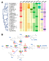
Figure 1
Figure 1
(A) Gene numbers of COX, PGSs, and PGDH in 20 species from different taxa; (B) presences of genes involved in the prostaglandin pathway from four major taxa.
Figure 2
Figure 2
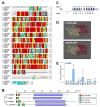
(A) Alignment of the COX a.a. sequences among four species including L. vannamei (XP027218437.1), H. sapiens (NP000954), D. magna (XP032795448.2), and C. gigas (XP011440168.2). The conserved residues are boxed in red, and similar residues are labeled in yellow; (B) the structural domains of COX proteins predicted by SMART; (C) exon/intron organization of LvCOX gene. The ORFs and UTRs in exons are marked in dark gray and empty boxes, respectively, and the introns are labeled with solid lines. The positions for 13 exons are 231–283, 2444–2557, 2723–2842, 2971–3114, 3307–3450, 3617–3798, 4019–4102, 4493–4633, 4943–5057, 5228–5403, 5561–5672, 5825–6038 and 6196–6444, and for 12 introns are 289–2443, 2558–2722, 2843–2970, 3115–3306, 3451–3616, 3799–4018, 4103–4492, 4634–4942, 5058–5227, 5404–5560, 5673–5824, and 6039–6195; (D) 3D structures and substrate binding regions of COXs in L. vannamei and H. sapiens; (E) relative mRNA ratio of LvCOX transcripts among different tissues including the brain (Br), eyestalk (Es), gills (Gi), hemocyte (He), hepatopancreas (Hp), heart (Ht), intestines (In), muscle (Ms), ovary (Ov), stomach (St), thoracic ganglion (Tn), testis (Ts), ventral ganglion (Vn). The experimental groups denoted by the same letter represent a similar level of transcript expression (p > 0.05, ANOVA, followed by Fisher LSD test).
Figure 3
Figure 3
(A) phylogenetic analysis and the structural domain organization of multiple PGS genes in various species. The selected species include D. magna, L. vannamei, H. sapiens, D. rerio, and Mus musculus; (B) relative mRNA ratio of multiple LvPGS genes (LvPGES2, LvcPGES, LvPGFS, LvAKR1C3, LvPGDS1, LvPGDS2, and LvTxA2S) among different tissues, including brain (Br), eyestalk (Es), gills (Gi), hemocyte (He), hepatopancreas (Hp), heart (Ht), intestines (In), muscle (Ms), ovary (Ov), stomach (St), thoracic ganglion (Tn), testis (Ts), and ventral ganglion (Vn). The experimental groups denoted by the same letter represent a similar level of transcript expression (p > 0.05, ANOVA, followed by Fisher LSD test).
Figure 4
Figure 4
(A) Alignment of PGDH a.a. sequences among four species (H. sapiens, X. tropicalis, C. gigas, and L. vannamei); (B) relative mRNA ratio of LvPGDH transcripts among different tissues including the brain (Br), eyestalk (Es), gills (Gi), hemocyte (He), hepatopancreas (Hp), heart (Ht), intestines (In), muscle (Ms), ovary (Ov), stomach (St), thoracic ganglion (Tn), testis (Ts), ventral ganglion (Vn). The experimental groups denoted by the same letter represent a similar level of transcript expression (p > 0.05, ANOVA, followed by Fisher LSD test).
Figure 5
Figure 5
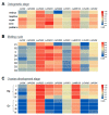
The expression profiles of genes involved in the PG pathway of L. vannamei during different developmental processes: (A) ontogenetic stages, including embryo, nauplius, mysis, zoea, and post-larvae; (B) molting cycle, including postmolt (P1 and P2 stages), intermolt (C stage) and premolt (D0, D1, D2, D3, and D4 stages); (C) ovarian development, including hepatopancreas (Hp) and ovaries (Ov) in stages I to IV.

Similar articles

Cited by

References

    1. Cha Y.I., Solnica-Krezel L., DuBois R.N. Fishing for prostanoids: Deciphering the developmental functions of cyclooxygenase-derived prostaglandins. Dev. Biol. 2006;289:263–272. doi: 10.1016/j.ydbio.2005.10.013. - DOI - PubMed
    1. Sugimoto Y., Narumiya S., Ichikawa A. Distribution and function of prostanoid receptors: Studies from knockout mice. Prog. Lipid Res. 2000;39:289–314. doi: 10.1016/S0163-7827(00)00008-4. - DOI - PubMed
    1. Funk C.D. Prostaglandins and leukotrienes: Advances in eicosanoid biology. Science. 2001;294:1871–1875. doi: 10.1126/science.294.5548.1871. - DOI - PubMed
    1. Hata A.N., Breyer R.M. Pharmacology and signaling of prostaglandin receptors: Multiple roles in inflammation and immune modulation. Pharmacol. Ther. 2004;103:147–166. doi: 10.1016/j.pharmthera.2004.06.003. - DOI - PubMed
    1. Christ E.J., van Dorp D.A. Comparative aspects of prostaglandin biosynthesis in animal tissues. Biochim. Biophys. Acta. 1972;270:534–545. doi: 10.1016/0005-2760(72)90119-1. - DOI - PubMed

LinkOut - more resources