Review
. 2022 Jan 31;23(3):1656.
doi: 10.3390/ijms23031656.

Broad Specific Xyloglucan:Xyloglucosyl Transferases Are Formidable Players in the Re-Modelling of Plant Cell Wall Structures

Affiliations
Review

Broad Specific Xyloglucan:Xyloglucosyl Transferases Are Formidable Players in the Re-Modelling of Plant Cell Wall Structures

Maria Hrmova et al. Int J Mol Sci. .

Abstract

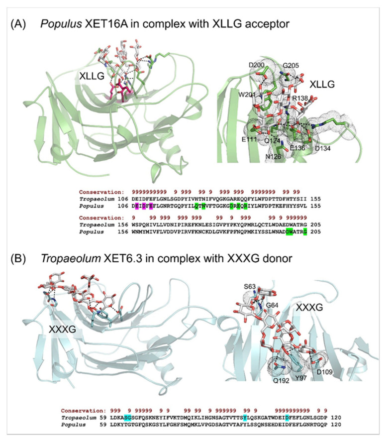
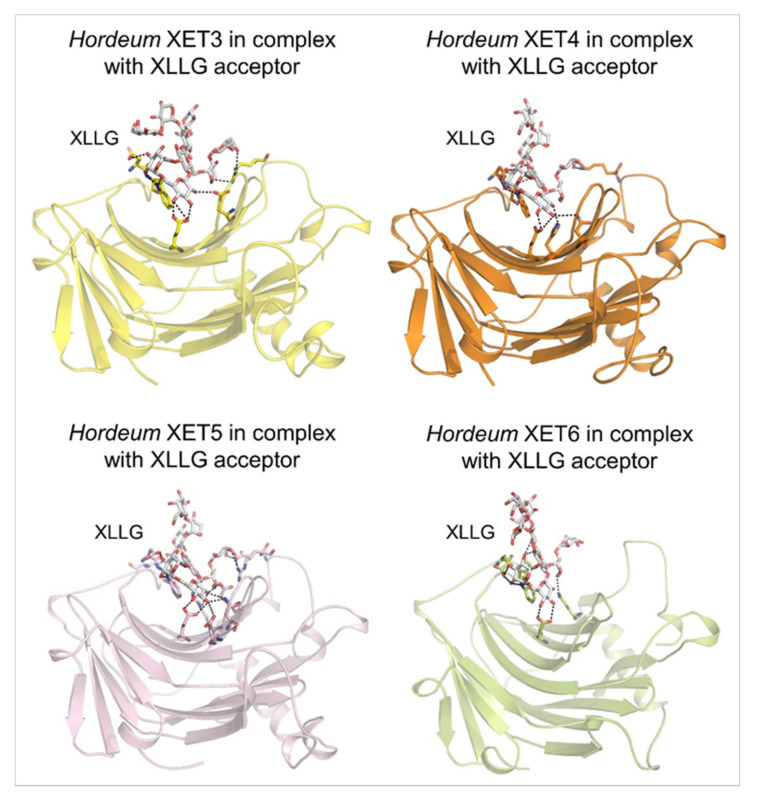
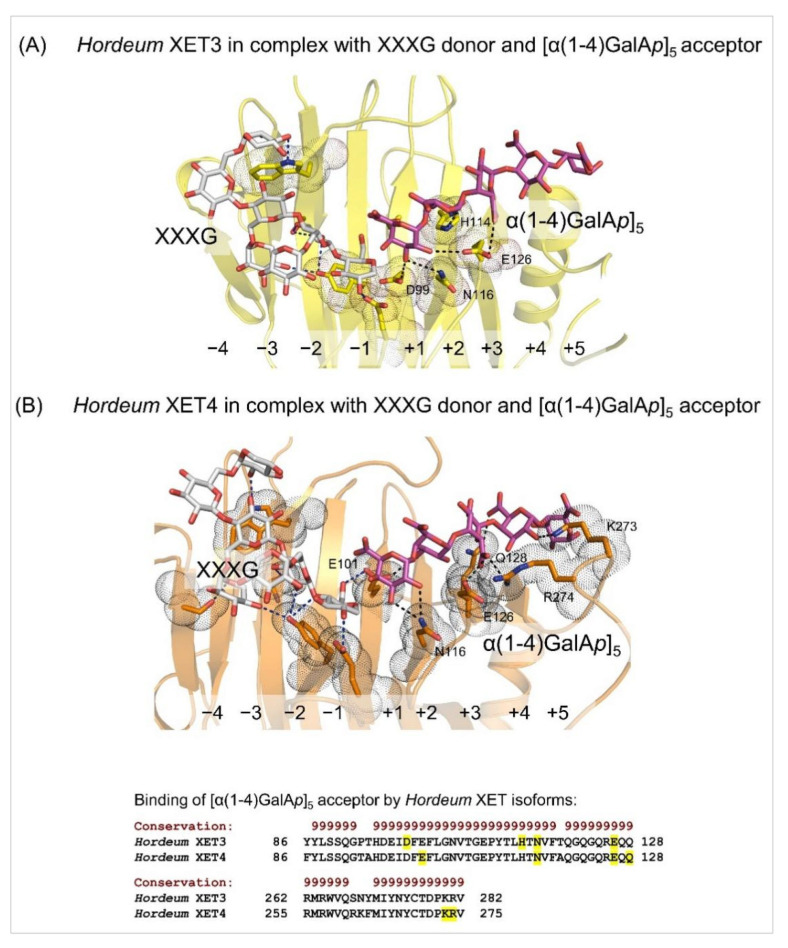
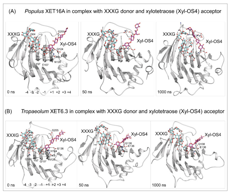
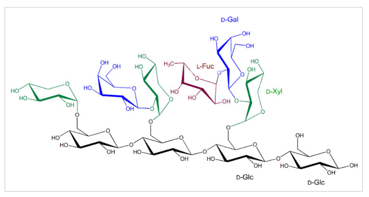
Plant xyloglucan:xyloglucosyl transferases, known as xyloglucan endo-transglycosylases (XETs) are the key players that underlie plant cell wall dynamics and mechanics. These fundamental roles are central for the assembly and modifications of cell walls during embryogenesis, vegetative and reproductive growth, and adaptations to living environments under biotic and abiotic (environmental) stresses. XET enzymes (EC 2.4.1.207) have the β-sandwich architecture and the β-jelly-roll topology, and are classified in the glycoside hydrolase family 16 based on their evolutionary history. XET enzymes catalyse transglycosylation reactions with xyloglucan (XG)-derived and other than XG-derived donors and acceptors, and this poly-specificity originates from the structural plasticity and evolutionary diversification that has evolved through expansion and duplication. In phyletic groups, XETs form the gene families that are differentially expressed in organs and tissues in time- and space-dependent manners, and in response to environmental conditions. Here, we examine higher plant XET enzymes and dissect how their exclusively carbohydrate-linked transglycosylation catalytic function inter-connects complex plant cell wall components. Further, we discuss progress in technologies that advance the knowledge of plant cell walls and how this knowledge defines the roles of XETs. We construe that the broad specificity of the plant XETs underscores their roles in continuous cell wall restructuring and re-modelling.

Keywords: crystal structures; evolutionary history; glycoside hydrolase family 16; mechanism of catalysis; molecular modelling and dynamics; substrate binding; transglycosylation reactions.


Conflict of interest statement

The authors declare no conflict of interest.

Figures

Figure 2
Figure 2
β-Sandwich architectures and β-jelly-roll topologies of Populus XET16A (PDB accession 1UMZ) in complex with the XLLG acceptor substrate (A), and the Tropaeolum XET6.3 3D molecular model in complex with the XXXG donor substrate (B). (A) Left panel: The XLLG acceptor (cpk sticks) bound in the Populus XET16A structure is indicated in dashed lines at 2.6 Å to 3.5 Å separations. The catalytic trio (Glu85, Glu89 and Asp87) is shown in cpk magenta sticks. Right panel: Details of the XLLG binding in Populus XET16A; interacting residues are marked in green cpk sticks and dots. Bottom panel: Some of the residues of Populues XET16A binding XLLG (highlighted in green) are shown in the alignment by PROMALS3D [83] of the Populus and Tropaeolum XET sequences (numbering includes signal peptides). Conservation of residues on the scale 9–6 is shown on the top of the alignment in brown. (B) Left panel: The XXXG donor (cpk sticks) bound in Tropaeolum XET6.3 (cyan; coordinates from [66]) is indicated in dashed lines at separations between 2.3 Å and 3.0 Å. Right panel: Details of XXXG binding in Tropaeolum XET6.3; interacting residues are marked in cyan cpk sticks in dots. Bottom panel: Some of the residues of Tropaeolum XET6.3 binding XLLG (highlighted in cyan) are shown in the alignment by PROMALS3D [83] of Populus XET16A and Tropaeolum XET6.3 sequences (numbering includes signal peptides). Conservation of residues on the scale 9–6 is shown on the top of the alignment in brown. Images were generated in the PyMOL Molecular Graphics System v2.5.2 (Schrődinger LLC, Portland, OR, USA).
Figure 3
Figure 3
Molecular models of the β-sandwich architecture with the β-jelly-roll topology of Hordeum XET3 (top left; yellow), Hordeum XET4 (top right; orange), Hordeum XET5 (bottom left; pink), and Hordeum XET6 (bottom right; lemon) in complex with the XLLG acceptors (cpk sticks). Coordinates of XET3, XET4 and XET6 with XLLG were taken from [81] and those of XET5 from [64]. XLLG was docked in XET5 by HDOCK that performs docking based on a hybrid algorithm of template-based modelling and ab initio free docking [98]; the top docking pose is shown for XET5 from 100 poses ranked by energy docking scores. Details of binding of XLLG by Hordeum XETs are shown in dashed lines. Separations are for XET3: 2.7 Å to 3.6 Å; XET4: 2.6 Å to 3.5 Å; XET5: 2.5 Å to 3.6 Å; XET6: 2.5 Å to 3.2 Å. Images were generated in PyMOL as referenced in Figure 2.
Figure 4
Figure 4
Details of the XXXG donor (cpk sticks) and the [α(1-4)GalAp]5 acceptor (magenta cpk sticks) binding substrates in Hordeum XET3 (A) and Hordeum XET4 (B). In both panels, blue and black dashed lines indicate residue separations between donors or acceptors that are 2.6 Å–3.6 Å (XET3) and 2.7 Å–3.4 Å (XET4). Interacting residues are shown in yellow (XET3) and orange (XET4) cpk sticks and emphasised in dots. Subsites at -4 to -1 for XXXG and +1 to +5 for [α(1-4)GalAp]5 are indicated. Images were generated in PyMOL as referenced in Figure 2. Bottom panel: Some of the residues of Hordeum XETs that bind [α(1-4)GalAp]5 (highlighted in yellow) are shown in the alignment by PROMALS3D [83] of the Hordeum XET sequences (numbering includes signal peptides). Conservation of residues on the scale 9–6 is shown on the top of the alignment in brown.
Figure 5
Figure 5
Docking of the XXXG donor (cpk cyan sticks) and the xylotetraose (Xyl-OS4) acceptor (cpk magenta sticks) substrates in the active sites of Populus XET16A (PDB accession 1UN1) (A) and Tropaeolum XET6.3 [66] (B), and MD simulations of enzyme/substrate complexes after 0, 50, and 1000 ns (simulation times indicated in bottom-left corners). MD simulations were carried out similarly as described in [103]. Separations between the donors and acceptor substrates are between 2.6 Å and 3.6 Å for Populus XET16A and 2.7 Å–3.4 Å for Tropaeolum XET6.3. Residues (numbering includes signal peptides) mediating contacts with substrates are shown in cpk sticks. In Tropaeolum XET6.3, E136 and Q138 stabilise the binding of the Xyl-OS4 acceptor. For clarity, the residues and the subsite binding sites (−4 to −1 for XXXG and +1 to +4 for Xyl-OS) are shown in left panels only. Images were generated in PyMOL as referenced in Figure 2.
Figure 1
Figure 1
The common substitutions in the tetragluco-oligosaccharide unit of XGs that forms chains of repeating (1,4)-β-d-linked glucopyranosyl residues (black), with the C-6 carbons carrying α-d-xylopyranosyl moieties (green), which could be substituted by galactopyranosyl residues (blue) on the C-2 carbons (β-d-Galp-(1,2)-α-d-Xylp). The galactopyranosyl residues could carry fucopyranosyl residues (brown) on the C-2 carbons (α-l-Fucp-(1,2)-β-d-Galp-(1,2)-α-d-Xylp). Descriptions of sugar moieties are indicated in matching colours.

Similar articles

Cited by

References

    1. Cosgrove D. Growth of the plant cell wall. Nat. Rev. Mol. Cell. Biol. 2005;6:850–861. doi: 10.1038/nrm1746. - DOI - PubMed
    1. Farrokhi N., Burton R.A., Brownfield L., Hrmova M., Wilson S.M., Bacic A., Fincher G.B. Plant cell wall biosynthesis: Genetic, biochemical and functional genomics approaches to the identification of key genes. Plant Biotech. J. 2009;4:145–167. doi: 10.1111/j.1467-7652.2005.00169.x. - DOI - PubMed
    1. Cosgrove D.J. Plant cell wall extensibility: Connecting plant cell growth with cell wall structure, mechanics, and the action of wall modifying enzymes. J. Exp. Bot. 2015;67:463–476. doi: 10.1093/jxb/erv511. - DOI - PubMed
    1. Popper Z.A., Michel G., Hervé C., Domozych D.S., Willats W.G., Tuohy M.G., Kloareg B., Stengel D.B. Evolution and diversity of plant cell walls: From algae to flowering plants. Annu. Rev. Plant Biol. 2011;62:567–590. doi: 10.1146/annurev-arplant-042110-103809. - DOI - PubMed
    1. Scheller H.V., Ulvskov P. Hemicelluloses. Annu. Rev. Plant Biol. 2010;61:263–289. doi: 10.1146/annurev-arplant-042809-112315. - DOI - PubMed

LinkOut - more resources